


4NTW
Structure of acid-sensing ion channel in complex with snake toxin
- PDB DOI: https://doi.org/10.2210/pdb4NTW/pdb
- Classification: TRANSPORT PROTEIN/TOXIN
- Organism(s): Gallus gallus, Micrurus tener tener
- Expression System: Homo sapiens, Escherichia coli
- Mutation(s): No
- Membrane Protein: Yes PDBTMMemProtMDmpstruc
- Deposited: 2013-12-02 Released: 2014-02-19
Experimental Data Snapshot
- Method: X-RAY DIFFRACTION
- Resolution: 2.07 Å
- R-Value Free: 0.246
- R-Value Work: 0.206
- R-Value Observed: 0.208

This is version 2.1 of the entry. See complete history.

Macromolecules
Find similar proteins by:
(by identity cutoff) | 3D Structure
Entity ID: 1 | |||||
|---|---|---|---|---|---|
| Molecule | Chains | Sequence Length | Organism | Details | Image |
| Acid-sensing ion channel 1 | 450 | Gallus gallus | Mutation(s): 0 Gene Names: ASIC1, ACCN2 Membrane Entity: Yes | 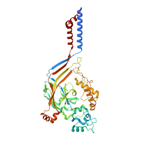 | |
UniProt | |||||
Find proteins for Q1XA76 (Gallus gallus) Explore Q1XA76 Go to UniProtKB: Q1XA76 | |||||
Entity Groups | |||||
| Sequence Clusters | 30% Identity50% Identity70% Identity90% Identity95% Identity100% Identity | ||||
| UniProt Group | Q1XA76 | ||||
Sequence AnnotationsExpand | |||||
| |||||
Find similar proteins by:
(by identity cutoff) | 3D Structure
Entity ID: 2 | |||||
|---|---|---|---|---|---|
| Molecule | Chains | Sequence Length | Organism | Details | Image |
| Neurotoxin MitTx-alpha | 60 | Micrurus tener tener | Mutation(s): 0 Membrane Entity: Yes | 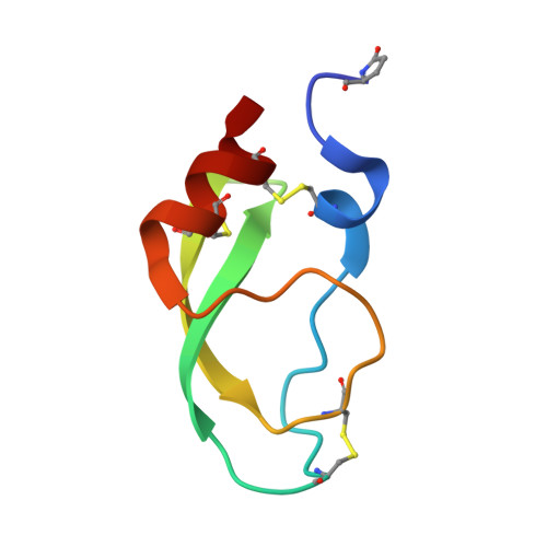 | |
UniProt | |||||
Find proteins for G9I929 (Micrurus tener tener) Explore G9I929 Go to UniProtKB: G9I929 | |||||
Entity Groups | |||||
| Sequence Clusters | 30% Identity50% Identity70% Identity90% Identity95% Identity100% Identity | ||||
| UniProt Group | G9I929 | ||||
Sequence AnnotationsExpand | |||||
| |||||
Find similar proteins by:
(by identity cutoff) | 3D Structure
Entity ID: 3 | |||||
|---|---|---|---|---|---|
| Molecule | Chains | Sequence Length | Organism | Details | Image |
| Basic phospholipase A2 homolog Tx-beta | 119 | Micrurus tener tener | Mutation(s): 0 Membrane Entity: Yes | 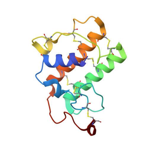 | |
UniProt | |||||
Find proteins for G9I930 (Micrurus tener tener) Explore G9I930 Go to UniProtKB: G9I930 | |||||
Entity Groups | |||||
| Sequence Clusters | 30% Identity50% Identity70% Identity90% Identity95% Identity100% Identity | ||||
| UniProt Group | G9I930 | ||||
Sequence AnnotationsExpand | |||||
| |||||
Small Molecules
| Ligands 4 Unique | |||||
|---|---|---|---|---|---|
| ID | Chains | Name / Formula / InChI Key | 2D Diagram | 3D Interactions | |
| P6G Query on P6G | I [auth A] | HEXAETHYLENE GLYCOL C12 H26 O7 IIRDTKBZINWQAW-UHFFFAOYSA-N |  | ||
| NAG Query on NAG | D [auth A], E [auth A] | 2-acetamido-2-deoxy-beta-D-glucopyranose C8 H15 N O6 OVRNDRQMDRJTHS-FMDGEEDCSA-N |  | ||
| CL Query on CL | F [auth A] | CHLORIDE ION Cl VEXZGXHMUGYJMC-UHFFFAOYSA-M |  | ||
| NA Query on NA | G [auth A], H [auth A], J [auth C] | SODIUM ION Na FKNQFGJONOIPTF-UHFFFAOYSA-N |  | ||
| Modified Residues 1 Unique | |||||
|---|---|---|---|---|---|
| ID | Chains | Type | Formula | 2D Diagram | Parent |
| PCA Query on PCA | B | L-PEPTIDE LINKING | C5 H7 N O3 |  | GLN |
Experimental Data & Validation
Experimental Data
- Method: X-RAY DIFFRACTION
- Resolution: 2.07 Å
- R-Value Free: 0.246
- R-Value Work: 0.206
- R-Value Observed: 0.208
- Space Group: H 3
Unit Cell:
| Length ( Å ) | Angle ( ˚ ) |
|---|---|
| a = 151.93 | α = 90 |
| b = 151.93 | β = 90 |
| c = 123.79 | γ = 120 |
| Software Name | Purpose |
|---|---|
| ADSC | data collection |
| PHASER | phasing |
| PHENIX | refinement |
| XDS | data reduction |
| XDS | data scaling |
Entry History
Deposition Data
- Released Date: 2014-02-19 Deposition Author(s): Baconguis, I., Bohlen, C.J., Goehring, A., Julius, D., Gouaux, E.
Revision History (Full details and data files)
- Version 1.0: 2014-02-19
Type: Initial release - Version 1.1: 2014-03-12
Changes: Database references - Version 1.2: 2017-11-08
Changes: Source and taxonomy - Version 2.0: 2019-12-25
Changes: Database references, Derived calculations, Polymer sequence - Version 2.1: 2020-07-29
Type: Remediation
Reason: Carbohydrate remediation
Changes: Data collection, Derived calculations, Structure summary
