4NQW

Structure of Mycobacterium tuberculosis extracytoplasmic function sigma factor SigK in complex with the cytosolic domain of its cognate anti-sigma factor RskA


Experimental Data Snapshot

  • Method: X-RAY DIFFRACTION
  • Resolution: 2.40 Å
  • R-Value Free: 0.260 
  • R-Value Work: 0.205 
  • R-Value Observed: 0.210 

wwPDB Validation   3D Report Full Report


This is version 1.1 of the entry. See complete history


Literature

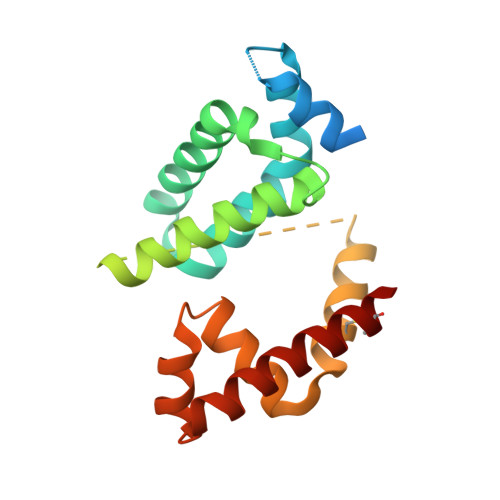
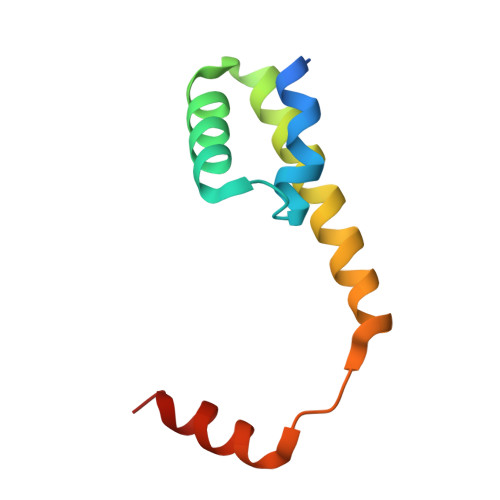
Structural basis for the redox sensitivity of the Mycobacterium tuberculosis SigK-RskA sigma-anti-sigma complex

Shukla, J.Gupta, R.Thakur, K.G.Gokhale, R.Gopal, B.

(2014) Acta Crystallogr D Biol Crystallogr 70: 1026-1036

  • DOI: https://doi.org/10.1107/S1399004714000121
  • Primary Citation of Related Structures:  
    4NQW

  • PubMed Abstract: 

    The host-pathogen interactions in Mycobacterium tuberculosis infection are significantly influenced by redox stimuli and alterations in the levels of secreted antigens. The extracytoplasmic function (ECF) σ factor σ(K) governs the transcription of the serodominant antigens MPT70 and MPT83. The cellular levels of σ(K) are regulated by the membrane-associated anti-σ(K) (RskA) that localizes σ(K) in an inactive complex. The crystal structure of M. tuberculosis σ(K) in complex with the cytosolic domain of RskA (RskAcyto) revealed a disulfide bridge in the -35 promoter-interaction region of σ(K). Biochemical experiments reveal that the redox potential of the disulfide-forming cysteines in σ(K) is consistent with its role as a sensor. The disulfide bond in σ(K) influences the stability of the σ(K)-RskAcyto complex but does not interfere with σ(K)-promoter DNA interactions. It is noted that these disulfide-forming cysteines are conserved across homologues, suggesting that this could be a general mechanism for redox-sensitive transcription regulation.


  • Organizational Affiliation

    Molecular Biophysics Unit, Indian Institute of Science, Bangalore 560 012, India.


Macromolecules
Find similar proteins by:  (by identity cutoff)  |  3D Structure
Entity ID: 1
MoleculeChains Sequence LengthOrganismDetailsImage
ECF RNA polymerase sigma factor SigK204Mycobacterium tuberculosisMutation(s): 0 
Gene Names: Rv0445csigK
UniProt
Find proteins for P9WGH7 (Mycobacterium tuberculosis (strain ATCC 25618 / H37Rv))
Explore P9WGH7 
Go to UniProtKB:  P9WGH7
Entity Groups  
Sequence Clusters30% Identity50% Identity70% Identity90% Identity95% Identity100% Identity
UniProt GroupP9WGH7
Sequence Annotations
Expand
  • Reference Sequence
Find similar proteins by:  (by identity cutoff)  |  3D Structure
Entity ID: 2
MoleculeChains Sequence LengthOrganismDetailsImage
Anti-sigma-K factor RskA80Mycobacterium tuberculosisMutation(s): 0 
Gene Names: rskARv0444c
UniProt
Find proteins for P9WGX5 (Mycobacterium tuberculosis (strain ATCC 25618 / H37Rv))
Explore P9WGX5 
Go to UniProtKB:  P9WGX5
Entity Groups  
Sequence Clusters30% Identity50% Identity70% Identity90% Identity95% Identity100% Identity
UniProt GroupP9WGX5
Sequence Annotations
Expand
  • Reference Sequence
Experimental Data & Validation

Experimental Data

  • Method: X-RAY DIFFRACTION
  • Resolution: 2.40 Å
  • R-Value Free: 0.260 
  • R-Value Work: 0.205 
  • R-Value Observed: 0.210 
  • Space Group: P 41 21 2
Unit Cell:
Length ( Å )Angle ( ˚ )
a = 57.39α = 90
b = 57.39β = 90
c = 150.87γ = 90
Software Package:
Software NamePurpose
MAR345dtbdata collection
SOLVEphasing
REFMACrefinement
MOSFLMdata reduction
SCALAdata scaling

Structure Validation

View Full Validation Report


Entry History 

Deposition Data

Revision History  (Full details and data files)

  • Version 1.0: 2014-01-22
    Type: Initial release
  • Version 1.1: 2014-04-16
    Changes: Database references