4NLI

Crystal structure of sheep beta-lactoglobulin (space group P3121)


Experimental Data Snapshot

  • Method: X-RAY DIFFRACTION
  • Resolution: 1.90 Å
  • R-Value Free: 0.206 
  • R-Value Work: 0.194 

wwPDB Validation   3D Report Full Report


This is version 1.4 of the entry. See complete history


Literature

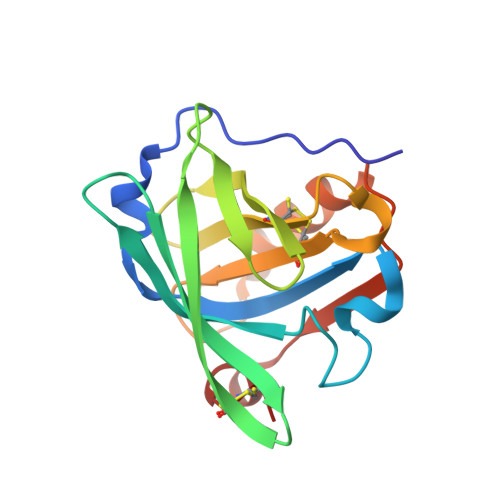
Structure of two crystal forms of sheep beta-lactoglobulin with EF-loop in closed conformation

Loch, J.I.Molenda, M.Kopec, M.Swiatek, S.Lewinski, K.

(2014) Biopolymers 101: 886-894

  • DOI: https://doi.org/10.1002/bip.22471
  • Primary Citation of Related Structures:  
    4NLI, 4NLJ

  • PubMed Abstract: 

    Ovine β-lactoglobulin has been isolated from whey fraction of sheep milk and crystallized. The high-resolution structures of two crystal forms (triclinic and trigonal) obtained at pH 7.0 have been determined revealing that ovine protein, similarly to its bovine analog, is dimeric. Access to the binding site located in the eight-stranded antiparallel β-barrel in both structures is blocked by the EF loop that has been found in closed conformation. Similarly to bovine lactoglobulin (BLG), conformation of the EF loop is stabilized by hydrogen bond between Glu89 and Ser116 indicating that Tanford transition might occur with the same mechanism. The substitution at six positions in relation to the most abundant isoform B of BLG also affects the distribution of electrostatic potentials and the total charge.


  • Organizational Affiliation

    Department of Crystal Chemistry and Crystal Physics, Faculty of Chemistry Jagiellonian University in Kraków, Ingardena 3, Kraków, 30-060, Poland.


Macromolecules
Find similar proteins by:  (by identity cutoff)  |  3D Structure
Entity ID: 1
MoleculeChains Sequence LengthOrganismDetailsImage
Beta-lactoglobulin-1/B162Ovis ariesMutation(s): 0 
UniProt
Find proteins for P67976 (Ovis aries)
Explore P67976 
Go to UniProtKB:  P67976
Entity Groups  
Sequence Clusters30% Identity50% Identity70% Identity90% Identity95% Identity100% Identity
UniProt GroupP67976
Sequence Annotations
Expand
  • Reference Sequence
Experimental Data & Validation

Experimental Data

  • Method: X-RAY DIFFRACTION
  • Resolution: 1.90 Å
  • R-Value Free: 0.206 
  • R-Value Work: 0.194 
  • Space Group: P 31 2 1
Unit Cell:
Length ( Å )Angle ( ˚ )
a = 97.44α = 90
b = 97.44β = 90
c = 68.24γ = 120
Software Package:
Software NamePurpose
MAR345data collection
PHASERphasing
REFMACrefinement
MOSFLMdata reduction
SCALAdata scaling

Structure Validation

View Full Validation Report


Entry History 

Deposition Data

Revision History  (Full details and data files)

  • Version 1.0: 2014-03-12
    Type: Initial release
  • Version 1.1: 2014-03-19
    Changes: Database references, Derived calculations
  • Version 1.2: 2014-12-10
    Changes: Database references
  • Version 1.3: 2017-11-22
    Changes: Data collection, Refinement description
  • Version 1.4: 2023-11-08
    Changes: Data collection, Database references, Derived calculations, Refinement description