4NI2

Crystal structure of the heterodimeric catalytic domain of wild-type human soluble guanylate cyclase


Experimental Data Snapshot

  • Method: X-RAY DIFFRACTION
  • Resolution: 1.90 Å
  • R-Value Free: 0.200 
  • R-Value Work: 0.160 
  • R-Value Observed: 0.162 

wwPDB Validation   3D Report Full Report


This is version 1.3 of the entry. See complete history


Literature

Interfacial residues promote an optimal alignment of the catalytic center in human soluble guanylate cyclase: heterodimerization is required but not sufficient for activity.

Seeger, F.Quintyn, R.Tanimoto, A.Williams, G.J.Tainer, J.A.Wysocki, V.H.Garcin, E.D.

(2014) Biochemistry 53: 2153-2165

  • DOI: https://doi.org/10.1021/bi500129k
  • Primary Citation of Related Structures:  
    4NI2

  • PubMed Abstract: 

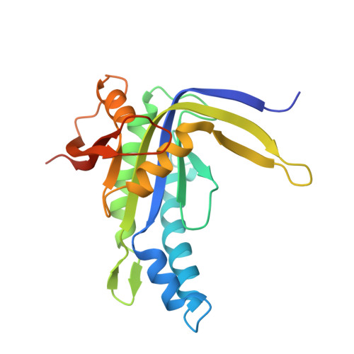
    Soluble guanylate cyclase (sGC) plays a central role in the cardiovascular system and is a drug target for the treatment of pulmonary hypertension. While the three-dimensional structure of sGC is unknown, studies suggest that binding of the regulatory domain to the catalytic domain maintains sGC in an autoinhibited basal state. The activation signal, binding of NO to heme, is thought to be transmitted via the regulatory and dimerization domains to the cyclase domain and unleashes the full catalytic potential of sGC. Consequently, isolated catalytic domains should show catalytic turnover comparable to that of activated sGC. Using X-ray crystallography, activity measurements, and native mass spectrometry, we show unambiguously that human isolated catalytic domains are much less active than basal sGC, while still forming heterodimers. We identified key structural elements regulating the dimer interface and propose a novel role for residues located in an interfacial flap and a hydrogen bond network as key modulators of the orientation of the catalytic subunits. We demonstrate that even in the absence of the regulatory domain, additional sGC domains are required to guide the appropriate conformation of the catalytic subunits associated with high activity. Our data support a novel regulatory mechanism whereby sGC activity is tuned by distinct domain interactions that either promote or inhibit catalytic activity. These results further our understanding of heterodimerization and activation of sGC and open additional drug discovery routes for targeting the NO-sGC-cGMP pathway via the design of small molecules that promote a productive conformation of the catalytic subunits or disrupt inhibitory domain interactions.


  • Organizational Affiliation

    University of Maryland Baltimore County , Baltimore, Maryland 21250, United States.


Macromolecules
Find similar proteins by:  (by identity cutoff)  |  3D Structure
Entity ID: 1
MoleculeChains Sequence LengthOrganismDetailsImage
Guanylate cyclase soluble subunit alpha-3197Homo sapiensMutation(s): 0 
Gene Names: GUC1A3GUCSA3GUCY1A1GUCY1A3
EC: 4.6.1.2
UniProt & NIH Common Fund Data Resources
Find proteins for Q02108 (Homo sapiens)
Explore Q02108 
Go to UniProtKB:  Q02108
PHAROS:  Q02108
GTEx:  ENSG00000164116 
Entity Groups  
Sequence Clusters30% Identity50% Identity70% Identity90% Identity95% Identity100% Identity
UniProt GroupQ02108
Sequence Annotations
Expand
  • Reference Sequence
Find similar proteins by:  (by identity cutoff)  |  3D Structure
Entity ID: 2
MoleculeChains Sequence LengthOrganismDetailsImage
Guanylate cyclase soluble subunit beta-1209Homo sapiensMutation(s): 0 
Gene Names: GUC1B3GUCSB3GUCY1B1GUCY1B3
EC: 4.6.1.2
UniProt & NIH Common Fund Data Resources
Find proteins for Q02153 (Homo sapiens)
Explore Q02153 
Go to UniProtKB:  Q02153
PHAROS:  Q02153
GTEx:  ENSG00000061918 
Entity Groups  
Sequence Clusters30% Identity50% Identity70% Identity90% Identity95% Identity100% Identity
UniProt GroupQ02153
Sequence Annotations
Expand
  • Reference Sequence
Experimental Data & Validation

Experimental Data

  • Method: X-RAY DIFFRACTION
  • Resolution: 1.90 Å
  • R-Value Free: 0.200 
  • R-Value Work: 0.160 
  • R-Value Observed: 0.162 
  • Space Group: P 21 21 21
Unit Cell:
Length ( Å )Angle ( ˚ )
a = 49.545α = 90
b = 55.834β = 90
c = 139.368γ = 90
Software Package:
Software NamePurpose
MOSFLMdata reduction
SCALAdata scaling
PHASERphasing
PHENIXrefinement
PDB_EXTRACTdata extraction
ELVESrefinement

Structure Validation

View Full Validation Report


Entry History 

Deposition Data

Revision History  (Full details and data files)

  • Version 1.0: 2014-04-16
    Type: Initial release
  • Version 1.1: 2014-05-07
    Changes: Database references
  • Version 1.2: 2017-11-22
    Changes: Refinement description
  • Version 1.3: 2023-09-20
    Changes: Data collection, Database references, Derived calculations, Refinement description