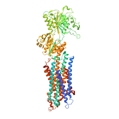
SERCA mutant E309Q binds two Ca(2+) ions but adopts a catalytically incompetent conformation.
Clausen, J.D., Bublitz, M., Arnou, B., Montigny, C., Jaxel, C., Moller, J.V., Nissen, P., Andersen, J.P., le Maire, M.(2013) EMBO J 32: 3231-3243
- PubMed: 24270570
- DOI: https://doi.org/10.1038/emboj.2013.250
- Primary Citation of Related Structures:
4NAB - PubMed Abstract:
The sarco(endo)plasmic reticulum Ca(2+)-ATPase (SERCA) couples ATP hydrolysis to transport of Ca(2+). This directed energy transfer requires cross-talk between the two Ca(2+) sites and the phosphorylation site over 50 Å distance. We have addressed the mechano-structural basis for this intramolecular signal by analysing the structure and the functional properties of SERCA mutant E309Q. Glu(309) contributes to Ca(2+) coordination at site II, and a consensus has been that E309Q only binds Ca(2+) at site I. The crystal structure of E309Q in the presence of Ca(2+) and an ATP analogue, however, reveals two occupied Ca(2+) sites of a non-catalytic Ca2E1 state. Ca(2+) is bound with micromolar affinity by both Ca(2+) sites in E309Q, but without cooperativity. The Ca(2+)-bound mutant does phosphorylate from ATP, but at a very low maximal rate. Phosphorylation depends on the correct positioning of the A-domain, requiring a shift of transmembrane segment M1 into an 'up and kinked position'. This transition is impaired in the E309Q mutant, most likely due to a lack of charge neutralization and altered hydrogen binding capacities at Ca(2+) site II.
Organizational Affiliation:
1] Department of Molecular Biology and Genetics, Centre for Membrane Pumps in Cells and Disease - PUMPKIN, Danish National Research Foundation, Aarhus University, Aarhus, Denmark [2] Department of Biomedicine, Aarhus University, Aarhus, Denmark.