4N8V

Crystal structure of killer cell immunoglobulin-like receptor KIR2DS2 in complex with HLA-A


Experimental Data Snapshot

  • Method: X-RAY DIFFRACTION
  • Resolution: 2.50 Å
  • R-Value Free: 0.257 
  • R-Value Work: 0.222 
  • R-Value Observed: 0.224 

wwPDB Validation   3D Report Full Report


This is version 1.3 of the entry. See complete history


Literature

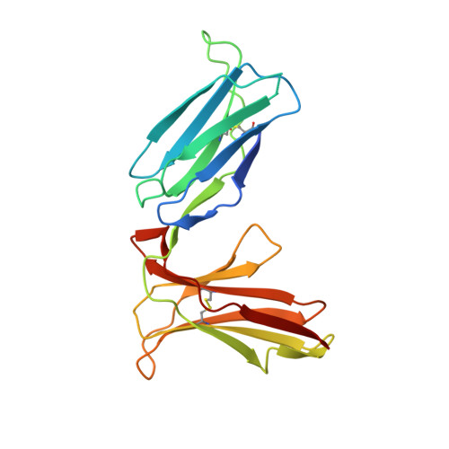
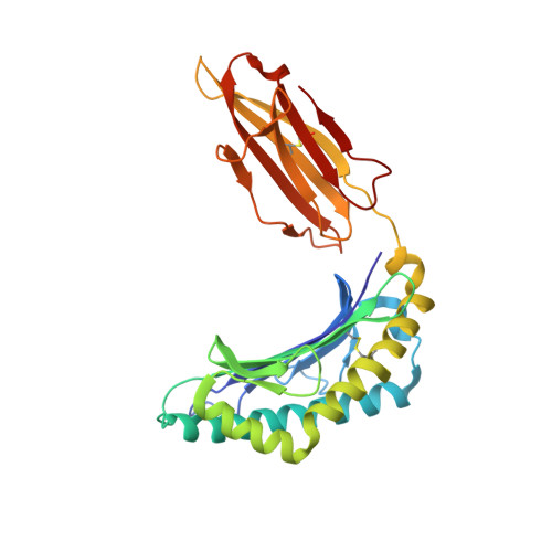
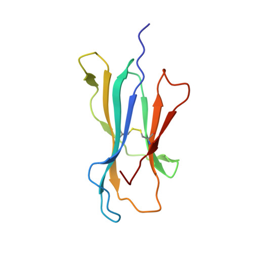
Activating killer cell immunoglobulin-like receptor 2DS2 binds to HLA-A*11

Liu, J.X.Xiao, Z.W.Ko, H.L.Shen, M.X.Ren, E.C.

(2014) Proc Natl Acad Sci U S A 111: 2662-2667

  • DOI: https://doi.org/10.1073/pnas.1322052111
  • Primary Citation of Related Structures:  
    4N8V

  • PubMed Abstract: 

    Inhibitory killer cell Ig-like receptors (KIRs) are known to recognize HLA ligands mainly of the HLA-C and Bw4 groups, but the ligands for KIRs are poorly understood. We report here the identification of the cognate ligand for the activating KIR 2DS2 as HLA-A*11:01. The crystal structure of the KIR2DS2-HLA-A*11:01 complex was solved at 2.5-Å resolution and revealed residue-binding characteristics distinct from those of inhibitory KIRs with HLA-C and the critical role of residues Tyr45 and Asp72 in shaping binding specificity to HLA-A*11:01. Using KIR2DS2 tetramers, binding to surface HLA-A*11:01 on live cells was demonstrated and, furthermore, that binding can be altered by residue changes at p8 of the peptide, indicating the influence of peptide sequence on KIR-HLA association. In addition, heteronuclear single quantum coherence NMR was used to map the involvement of critical residues in HLA binding at the interface of KIR and HLA, and validates the data observed in the crystal structure. Our data provide structural evidence of the recognition of A*11:01 by the activating KIR2DS2 and extend our understanding of the KIR-HLA binding spectrum.


  • Organizational Affiliation

    Singapore Immunology Network, Agency for Science, Technology and Research, Singapore 138648.


Macromolecules
Find similar proteins by:  (by identity cutoff)  |  3D Structure
Entity ID: 1
MoleculeChains Sequence LengthOrganismDetailsImage
Killer cell immunoglobulin-like receptor 2DS2A [auth G],
B [auth I]
201Homo sapiensMutation(s): 0 
Gene Names: KIR2DS2
UniProt
Find proteins for P43631 (Homo sapiens)
Explore P43631 
Go to UniProtKB:  P43631
Entity Groups  
Sequence Clusters30% Identity50% Identity70% Identity90% Identity95% Identity100% Identity
UniProt GroupP43631
Sequence Annotations
Expand
  • Reference Sequence
Find similar proteins by:  (by identity cutoff)  |  3D Structure
Entity ID: 2
MoleculeChains Sequence LengthOrganismDetailsImage
HLA class I histocompatibility antigen, A-11 alpha chainC [auth A],
F [auth D]
274Homo sapiensMutation(s): 0 
Gene Names: HLA-A
UniProt & NIH Common Fund Data Resources
Find proteins for P04439 (Homo sapiens)
Explore P04439 
Go to UniProtKB:  P04439
PHAROS:  P04439
GTEx:  ENSG00000206503 
Entity Groups  
Sequence Clusters30% Identity50% Identity70% Identity90% Identity95% Identity100% Identity
UniProt GroupP04439
Sequence Annotations
Expand
  • Reference Sequence
Find similar proteins by:  (by identity cutoff)  |  3D Structure
Entity ID: 3
MoleculeChains Sequence LengthOrganismDetailsImage
Beta-2-microglobulinD [auth B],
G [auth E]
100Homo sapiensMutation(s): 0 
Gene Names: B2M
UniProt & NIH Common Fund Data Resources
Find proteins for P61769 (Homo sapiens)
Explore P61769 
Go to UniProtKB:  P61769
PHAROS:  P61769
GTEx:  ENSG00000166710 
Entity Groups  
Sequence Clusters30% Identity50% Identity70% Identity90% Identity95% Identity100% Identity
UniProt GroupP61769
Sequence Annotations
Expand
  • Reference Sequence

Find similar proteins by:  Sequence   |   3D Structure  

Entity ID: 4
MoleculeChains Sequence LengthOrganismDetailsImage
peptide from Virion membrane protein A14E [auth C],
H [auth F]
9Vaccinia virus WRMutation(s): 0 
UniProt
Find proteins for Q76ZQ3 (Vaccinia virus (strain Western Reserve))
Explore Q76ZQ3 
Go to UniProtKB:  Q76ZQ3
Entity Groups  
UniProt GroupQ76ZQ3
Sequence Annotations
Expand
  • Reference Sequence
Experimental Data & Validation

Experimental Data

  • Method: X-RAY DIFFRACTION
  • Resolution: 2.50 Å
  • R-Value Free: 0.257 
  • R-Value Work: 0.222 
  • R-Value Observed: 0.224 
  • Space Group: P 21 21 21
Unit Cell:
Length ( Å )Angle ( ˚ )
a = 69.55α = 90
b = 94.67β = 90
c = 228.44γ = 90
Software Package:
Software NamePurpose
HKL-2000data collection
AMoREphasing
REFMACrefinement
MOSFLMdata reduction
SCALAdata scaling

Structure Validation

View Full Validation Report


Entry History 

Deposition Data

Revision History  (Full details and data files)

  • Version 1.0: 2014-02-05
    Type: Initial release
  • Version 1.1: 2014-02-12
    Changes: Database references
  • Version 1.2: 2014-03-12
    Changes: Database references
  • Version 1.3: 2023-11-08
    Changes: Data collection, Database references, Refinement description