


Crystal structure of phosphorylated phosphopentomutase from streptococcus mutans
Fedorov, A.A., Fedorov, E.V., Bonanno, J., Burley, S.K., Almo, S.C.To be published.
Experimental Data Snapshot
wwPDB Validation 3D Report Full Report

Entity ID: 1 | |||||
|---|---|---|---|---|---|
| Molecule | Chains | Sequence Length | Organism | Details | Image |
| Phosphopentomutase | 413 | Streptococcus mutans UA159 | Mutation(s): 0 Gene Names: deoB, SMU_1233 EC: 5.4.2.7 | 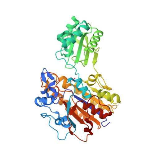 | |
UniProt | |||||
Find proteins for Q8DTU0 (Streptococcus mutans serotype c (strain ATCC 700610 / UA159)) Explore Q8DTU0 Go to UniProtKB: Q8DTU0 | |||||
Entity Groups | |||||
| Sequence Clusters | 30% Identity50% Identity70% Identity90% Identity95% Identity100% Identity | ||||
| UniProt Group | Q8DTU0 | ||||
Sequence AnnotationsExpand | |||||
| |||||
| Ligands 5 Unique | |||||
|---|---|---|---|---|---|
| ID | Chains | Name / Formula / InChI Key | 2D Diagram | 3D Interactions | |
| PEG Query on PEG | I [auth A] | DI(HYDROXYETHYL)ETHER C4 H10 O3 MTHSVFCYNBDYFN-UHFFFAOYSA-N |  | ||
| SO4 Query on SO4 | E [auth A], M [auth B] | SULFATE ION O4 S QAOWNCQODCNURD-UHFFFAOYSA-L |  | ||
| GOL Query on GOL | F [auth A], G [auth A], J [auth A] | GLYCEROL C3 H8 O3 PEDCQBHIVMGVHV-UHFFFAOYSA-N |  | ||
| MN Query on MN | C [auth A], D [auth A], K [auth B], L [auth B] | MANGANESE (II) ION Mn WAEMQWOKJMHJLA-UHFFFAOYSA-N |  | ||
| AZI Query on AZI | H [auth A], N [auth B] | AZIDE ION N3 IVRMZWNICZWHMI-UHFFFAOYSA-N |  | ||
| Modified Residues 1 Unique | |||||
|---|---|---|---|---|---|
| ID | Chains | Type | Formula | 2D Diagram | Parent |
| TPO Query on TPO | A, B | L-PEPTIDE LINKING | C4 H10 N O6 P |  | THR |
| Length ( Å ) | Angle ( ˚ ) |
|---|---|
| a = 69.788 | α = 90 |
| b = 63.687 | β = 98.85 |
| c = 104.765 | γ = 90 |
| Software Name | Purpose |
|---|---|
| ADSC | data collection |
| BALBES | phasing |
| PHENIX | refinement |
| HKL-2000 | data reduction |
| HKL-2000 | data scaling |
RCSB PDB (citation) is hosted by
RCSB PDB is a member of the

