4N07

Crystal structure of the GluA2 ligand-binding domain (S1S2J-L483Y-N754S) in complex with glutamate and BPAM-344 at 1.87 A resolution


Experimental Data Snapshot

  • Method: X-RAY DIFFRACTION
  • Resolution: 1.87 Å
  • R-Value Free: 0.211 
  • R-Value Work: 0.175 
  • R-Value Observed: 0.177 

wwPDB Validation   3D Report Full Report


Ligand Structure Quality Assessment 


This is version 1.3 of the entry. See complete history


Literature

Synthesis, pharmacological and structural characterization, and thermodynamic aspects of GluA2-positive allosteric modulators with a 3,4-dihydro-2H-1,2,4-benzothiadiazine 1,1-dioxide scaffold.

Nrholm, A.B.Francotte, P.Olsen, L.Krintel, C.Frydenvang, K.Goffin, E.Challal, S.Danober, L.Botez-Pop, I.Lestage, P.Pirotte, B.Kastrup, J.S.

(2013) J Med Chem 56: 8736-8745

  • DOI: https://doi.org/10.1021/jm4012092
  • Primary Citation of Related Structures:  
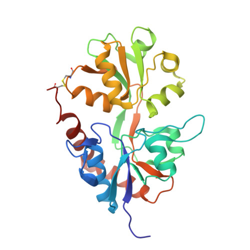
    4N07

  • PubMed Abstract: 

    Positive allosteric modulators of ionotropic glutamate receptors are potential compounds for treatment of cognitive disorders, e.g., Alzheimer's disease. The modulators bind within the dimer interface of the ligand-binding domain (LBD) and stabilize the agonist-bound conformation, thereby slowing receptor desensitization and/or deactivation. Here we describe the synthesis and pharmacological testing at GluA2 of a new generation of 3,4-dihydro-2H-1,2,4-benzothiadiazine 1,1-dioxides. The most potent modulator 3 in complex with GluA2-LBD-L483Y-N754S was subjected to structural analysis by X-ray crystallography, and the thermodynamics of binding was studied by isothermal titration calorimetry. Compound 3 binds to GluA2-LBD-L483Y-N754S with a Kd of 0.35 μM (ΔH = -7.5 kcal/mol and -TΔS = -1.3 kcal/mol). This is the first time that submicromolar binding affinity has been achieved for this type of positive allosteric modulator. The major structural factor increasing the binding affinity of 3 seems to be interactions between the cyclopropyl group of 3 and the backbone of Phe495 and Met496.


  • Organizational Affiliation

    Department of Drug Design and Pharmacology, Faculty of Health and Medical Sciences, University of Copenhagen , Universitetsparken 2, DK-2100 Copenhagen, Denmark.


Macromolecules
Find similar proteins by:  (by identity cutoff)  |  3D Structure
Entity ID: 1
MoleculeChains Sequence LengthOrganismDetailsImage
Glutamate receptor 2
A, B, C
263Rattus norvegicusMutation(s): 2 
Gene Names: Glur2Gria2
UniProt
Find proteins for P19491 (Rattus norvegicus)
Explore P19491 
Go to UniProtKB:  P19491
Entity Groups  
Sequence Clusters30% Identity50% Identity70% Identity90% Identity95% Identity100% Identity
UniProt GroupP19491
Sequence Annotations
Expand
  • Reference Sequence
Small Molecules
Ligands 6 Unique
IDChains Name / Formula / InChI Key2D Diagram3D Interactions
2J9
Query on 2J9

Download Ideal Coordinates CCD File 
D [auth A],
DA [auth C],
U [auth B]
4-cyclopropyl-7-fluoro-3,4-dihydro-2H-1,2,4-benzothiadiazine 1,1-dioxide
C10 H11 F N2 O2 S
FLTMTBPCYAZIKM-UHFFFAOYSA-N
GLU
Query on GLU

Download Ideal Coordinates CCD File 
E [auth A],
EA [auth C],
V [auth B]
GLUTAMIC ACID
C5 H9 N O4
WHUUTDBJXJRKMK-VKHMYHEASA-N
CAC
Query on CAC

Download Ideal Coordinates CCD File 
P [auth A]CACODYLATE ION
C2 H6 As O2
OGGXGZAMXPVRFZ-UHFFFAOYSA-M
GOL
Query on GOL

Download Ideal Coordinates CCD File 
BA [auth B]
CA [auth B]
Q [auth A]
R [auth A]
S [auth A]
BA [auth B],
CA [auth B],
Q [auth A],
R [auth A],
S [auth A],
T [auth A]
GLYCEROL
C3 H8 O3
PEDCQBHIVMGVHV-UHFFFAOYSA-N
ZN
Query on ZN

Download Ideal Coordinates CCD File 
AA [auth B]
KA [auth C]
L [auth A]
LA [auth C]
M [auth A]
AA [auth B],
KA [auth C],
L [auth A],
LA [auth C],
M [auth A],
MA [auth C],
N [auth A],
O [auth A],
Y [auth B],
Z [auth B]
ZINC ION
Zn
PTFCDOFLOPIGGS-UHFFFAOYSA-N
ACT
Query on ACT

Download Ideal Coordinates CCD File 
F [auth A]
FA [auth C]
G [auth A]
GA [auth C]
H [auth A]
F [auth A],
FA [auth C],
G [auth A],
GA [auth C],
H [auth A],
HA [auth C],
I [auth A],
IA [auth C],
J [auth A],
JA [auth C],
K [auth A],
W [auth B],
X [auth B]
ACETATE ION
C2 H3 O2
QTBSBXVTEAMEQO-UHFFFAOYSA-M
Binding Affinity Annotations 
IDSourceBinding Affinity
2J9 PDBBind:  4N07 Kd: 350 (nM) from 1 assay(s)
BindingDB:  4N07 Kd: 350 (nM) from 1 assay(s)
EC50: min: 810, max: 900 (nM) from 3 assay(s)
Binding MOAD:  4N07 Kd: 350 (nM) from 1 assay(s)
Experimental Data & Validation

Experimental Data

  • Method: X-RAY DIFFRACTION
  • Resolution: 1.87 Å
  • R-Value Free: 0.211 
  • R-Value Work: 0.175 
  • R-Value Observed: 0.177 
  • Space Group: P 21 21 2
Unit Cell:
Length ( Å )Angle ( ˚ )
a = 113.904α = 90
b = 164.034β = 90
c = 47.284γ = 90
Software Package:
Software NamePurpose
SCALAdata scaling
PHENIXrefinement
PDB_EXTRACTdata extraction
XDSdata reduction
PHASERphasing

Structure Validation

View Full Validation Report


Ligand Structure Quality Assessment 


Entry History 

Deposition Data

Revision History  (Full details and data files)

  • Version 1.0: 2013-11-20
    Type: Initial release
  • Version 1.1: 2014-03-19
    Changes: Database references
  • Version 1.2: 2017-08-09
    Changes: Data collection, Refinement description, Source and taxonomy
  • Version 1.3: 2023-09-20
    Changes: Data collection, Database references, Derived calculations, Refinement description