4MZU

Crystal structure of FdtD, a bifunctional ketoisomerase/N-acetyltransferase from Shewanella denitrificans


Experimental Data Snapshot

  • Method: X-RAY DIFFRACTION
  • Resolution: 2.20 Å
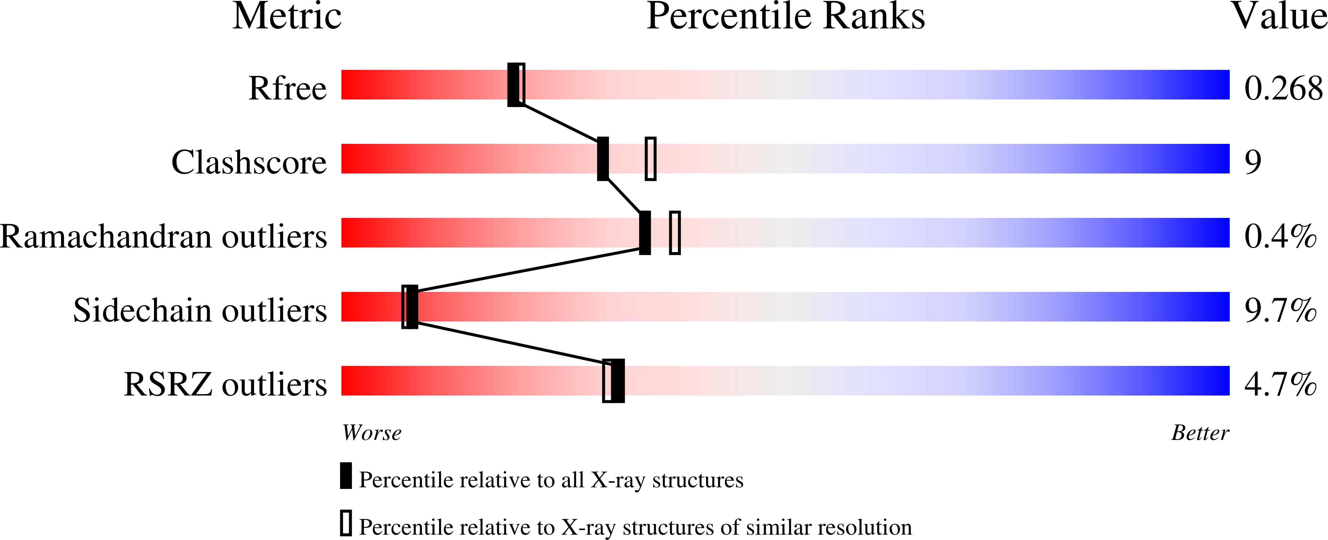
  • R-Value Free: 0.270 
  • R-Value Work: 0.204 
  • R-Value Observed: 0.208 

wwPDB Validation   3D Report Full Report


Ligand Structure Quality Assessment 


This is version 1.3 of the entry. See complete history


Literature

Structural and Biochemical Characterization of a Bifunctional Ketoisomerase/N-Acetyltransferase from Shewanella denitrificans.

Chantigian, D.P.Thoden, J.B.Holden, H.M.

(2013) Biochemistry 52: 8374-8385

  • DOI: https://doi.org/10.1021/bi401170t
  • Primary Citation of Related Structures:  
    4MZU

  • PubMed Abstract: 

    Unusual N-acetylated sugars have been observed on the O-antigens of some Gram-negative bacteria and on the S-layers of both Gram-positive and Gram-negative bacteria. One such sugar is 3-acetamido-3,6-dideoxy-α-d-galactose or Fuc3NAc. The pathway for its production requires five enzymes with the first step involving the attachment of dTMP to glucose-1-phosphate. Here, we report a structural and biochemical characterization of a bifunctional enzyme from Shewanella denitificans thought to be involved in the biosynthesis of dTDP-Fuc3NAc. On the basis of a bioinformatics analysis, the enzyme, hereafter referred to as FdtD, has been postulated to catalyze the third and fifth steps in the pathway, namely, a 3,4-keto isomerization and an N-acetyltransferase reaction. For the X-ray analysis reported here, the enzyme was crystallized in the presence of dTDP and CoA. The crystal structure shows that FdtD adopts a hexameric quaternary structure with 322 symmetry. Each subunit of the hexamer folds into two distinct domains connected by a flexible loop. The N-terminal domain adopts a left-handed β-helix motif and is responsible for the N-acetylation reaction. The C-terminal domain folds into an antiparallel flattened β-barrel that harbors the active site responsible for the isomerization reaction. Biochemical assays verify the two proposed catalytic activities of the enzyme and reveal that the 3,4-keto isomerization event leads to the inversion of configuration about the hexose C-4' carbon.


  • Organizational Affiliation

    Department of Biochemistry, University of Wisconsin , Madison, Wisconsin 53706, United States.


Macromolecules
Find similar proteins by:  (by identity cutoff)  |  3D Structure
Entity ID: 1
MoleculeChains Sequence LengthOrganismDetailsImage
WxcM-like protein
A, B, C, D, E
A, B, C, D, E, F, G, H, I, J, K, L
312Shewanella denitrificans OS217Mutation(s): 0 
Gene Names: FdtDSden_2659
UniProt
Find proteins for Q12KT8 (Shewanella denitrificans (strain OS217 / ATCC BAA-1090 / DSM 15013))
Explore Q12KT8 
Go to UniProtKB:  Q12KT8
Entity Groups  
Sequence Clusters30% Identity50% Identity70% Identity90% Identity95% Identity100% Identity
UniProt GroupQ12KT8
Sequence Annotations
Expand
  • Reference Sequence
Small Molecules
Ligands 4 Unique
IDChains Name / Formula / InChI Key2D Diagram3D Interactions
COA
Query on COA

Download Ideal Coordinates CCD File 
BA [auth E]
EA [auth F]
HA [auth G]
JA [auth G]
M [auth A]
BA [auth E],
EA [auth F],
HA [auth G],
JA [auth G],
M [auth A],
MA [auth I],
OA [auth J],
P [auth B],
SA [auth K],
T [auth C],
VA [auth L],
X [auth D]
COENZYME A
C21 H36 N7 O16 P3 S
RGJOEKWQDUBAIZ-IBOSZNHHSA-N
TYD
Query on TYD

Download Ideal Coordinates CCD File 
CA [auth E]
FA [auth F]
IA [auth G]
LA [auth H]
N [auth A]
CA [auth E],
FA [auth F],
IA [auth G],
LA [auth H],
N [auth A],
NA [auth I],
PA [auth J],
Q [auth B],
TA [auth K],
U [auth C],
WA [auth L],
Y [auth D]
THYMIDINE-5'-DIPHOSPHATE
C10 H16 N2 O11 P2
UJLXYODCHAELLY-XLPZGREQSA-N
TDR
Query on TDR

Download Ideal Coordinates CCD File 
DA [auth E]
GA [auth F]
O [auth A]
QA [auth J]
R [auth B]
DA [auth E],
GA [auth F],
O [auth A],
QA [auth J],
R [auth B],
UA [auth K],
V [auth C],
XA [auth L],
Z [auth D]
THYMINE
C5 H6 N2 O2
RWQNBRDOKXIBIV-UHFFFAOYSA-N
MG
Query on MG

Download Ideal Coordinates CCD File 
AA [auth D],
KA [auth G],
RA [auth J],
S [auth B],
W [auth C]
MAGNESIUM ION
Mg
JLVVSXFLKOJNIY-UHFFFAOYSA-N
Experimental Data & Validation

Experimental Data

  • Method: X-RAY DIFFRACTION
  • Resolution: 2.20 Å
  • R-Value Free: 0.270 
  • R-Value Work: 0.204 
  • R-Value Observed: 0.208 
  • Space Group: P 1
Unit Cell:
Length ( Å )Angle ( ˚ )
a = 85.314α = 79.23
b = 109.443β = 79.98
c = 127.847γ = 84.89
Software Package:
Software NamePurpose
HKL-3000data collection
SOLVEphasing
REFMACrefinement
HKL-3000data reduction
HKL-3000data scaling

Structure Validation

View Full Validation Report


Ligand Structure Quality Assessment 


Entry History 

Deposition Data

Revision History  (Full details and data files)

  • Version 1.0: 2013-10-09
    Type: Initial release
  • Version 1.1: 2013-12-18
    Changes: Database references
  • Version 1.2: 2024-02-28
    Changes: Data collection, Database references, Derived calculations
  • Version 1.3: 2024-04-03
    Changes: Refinement description