4MZE

Crystal structure of hPIV3 hemagglutinin-neuraminidase, H552Q/Q559R mutant


Experimental Data Snapshot

  • Method: X-RAY DIFFRACTION
  • Resolution: 1.80 Å
  • R-Value Free: 0.196 
  • R-Value Work: 0.161 
  • R-Value Observed: 0.163 

wwPDB Validation   3D Report Full Report


Ligand Structure Quality Assessment 


This is version 2.0 of the entry. See complete history


Literature

Interaction between the hemagglutinin-neuraminidase and fusion glycoproteins of human parainfluenza virus type III regulates viral growth in vivo.

Xu, R.Palmer, S.G.Porotto, M.Palermo, L.M.Niewiesk, S.Wilson, I.A.Moscona, A.

(2013) mBio 4: e00803-e00813

  • DOI: https://doi.org/10.1128/mBio.00803-13
  • Primary Citation of Related Structures:  
    4MZA, 4MZE

  • PubMed Abstract: 

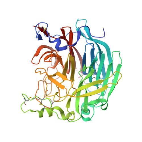
    Paramyxoviruses, enveloped RNA viruses that include human parainfluenza virus type 3 (HPIV3), cause the majority of childhood viral pneumonia. HPIV3 infection starts when the viral receptor-binding protein engages sialic acid receptors in the lung and the viral envelope fuses with the target cell membrane. Fusion/entry requires interaction between two viral surface glycoproteins: tetrameric hemagglutinin-neuraminidase (HN) and fusion protein (F). In this report, we define structural correlates of the HN features that permit infection in vivo. We have shown that viruses with an HN-F that promotes growth in cultured immortalized cells are impaired in differentiated human airway epithelial cell cultures (HAE) and in vivo and evolve in HAE into viable viruses with less fusogenic HN-F. In this report, we identify specific structural features of the HN dimer interface that modulate HN-F interaction and fusion triggering and directly impact infection. Crystal structures of HN, which promotes viral growth in vivo, show a diminished interface in the HN dimer compared to the reference strain's HN, consistent with biochemical and biological data indicating decreased dimerization and decreased interaction with F protein. The crystallographic data suggest a structural explanation for the HN's altered ability to activate F and reveal properties that are critical for infection in vivo.


  • Organizational Affiliation

    Department of Integrative Structural and Computational Biology, The Scripps Research Institute, La Jolla, California, USA.


Macromolecules
Find similar proteins by:  (by identity cutoff)  |  3D Structure
Entity ID: 1
MoleculeChains Sequence LengthOrganismDetailsImage
Hemagglutinin-neuraminidase
A, B
437Human parainfluenza 3 virus (strain NIH 47885)Mutation(s): 2 
Gene Names: HN
EC: 3.2.1.18
UniProt
Find proteins for P08492 (Human parainfluenza 3 virus (strain Wash/47885/57))
Explore P08492 
Go to UniProtKB:  P08492
Entity Groups  
Sequence Clusters30% Identity50% Identity70% Identity90% Identity95% Identity100% Identity
UniProt GroupP08492
Sequence Annotations
Expand
  • Reference Sequence
Oligosaccharides

Help

Entity ID: 2
MoleculeChains Length2D Diagram Glycosylation3D Interactions
alpha-D-mannopyranose-(1-2)-alpha-D-mannopyranose-(1-3)-[alpha-D-mannopyranose-(1-3)-[alpha-D-mannopyranose-(1-6)]alpha-D-mannopyranose-(1-6)]beta-D-mannopyranose-(1-4)-2-acetamido-2-deoxy-beta-D-glucopyranose-(1-4)-2-acetamido-2-deoxy-beta-D-glucopyranose
C
8N-Glycosylation
Glycosylation Resources
GlyTouCan:  G80966KZ
GlyCosmos:  G80966KZ
GlyGen:  G80966KZ
Entity ID: 3
MoleculeChains Length2D Diagram Glycosylation3D Interactions
beta-L-fucopyranose-(1-6)-2-acetamido-2-deoxy-beta-D-glucopyranose
D
2N-Glycosylation
Glycosylation Resources
GlyTouCan:  G61843VN
GlyCosmos:  G61843VN
GlyGen:  G61843VN
Entity ID: 4
MoleculeChains Length2D Diagram Glycosylation3D Interactions
alpha-D-mannopyranose-(1-3)-[alpha-D-mannopyranose-(1-6)]beta-D-mannopyranose-(1-4)-2-acetamido-2-deoxy-beta-D-glucopyranose-(1-4)-2-acetamido-2-deoxy-beta-D-glucopyranose
E
5N-Glycosylation
Glycosylation Resources
GlyTouCan:  G22768VO
GlyCosmos:  G22768VO
GlyGen:  G22768VO
Entity ID: 5
MoleculeChains Length2D Diagram Glycosylation3D Interactions
beta-D-mannopyranose-(1-4)-2-acetamido-2-deoxy-beta-D-glucopyranose-(1-4)-2-acetamido-2-deoxy-beta-D-glucopyranose
F
3N-Glycosylation
Glycosylation Resources
GlyTouCan:  G15407YE
GlyCosmos:  G15407YE
GlyGen:  G15407YE
Small Molecules
Ligands 6 Unique
IDChains Name / Formula / InChI Key2D Diagram3D Interactions
NAG
Query on NAG

Download Ideal Coordinates CCD File 
G [auth A],
U [auth B]
2-acetamido-2-deoxy-beta-D-glucopyranose
C8 H15 N O6
OVRNDRQMDRJTHS-FMDGEEDCSA-N
PEG
Query on PEG

Download Ideal Coordinates CCD File 
FA [auth B],
GA [auth B],
R [auth A],
S [auth A]
DI(HYDROXYETHYL)ETHER
C4 H10 O3
MTHSVFCYNBDYFN-UHFFFAOYSA-N
SO4
Query on SO4

Download Ideal Coordinates CCD File 
T [auth A]SULFATE ION
O4 S
QAOWNCQODCNURD-UHFFFAOYSA-L
PO4
Query on PO4

Download Ideal Coordinates CCD File 
H [auth A],
V [auth B],
W [auth B]
PHOSPHATE ION
O4 P
NBIIXXVUZAFLBC-UHFFFAOYSA-K
EDO
Query on EDO

Download Ideal Coordinates CCD File 
AA [auth B]
BA [auth B]
CA [auth B]
DA [auth B]
EA [auth B]
AA [auth B],
BA [auth B],
CA [auth B],
DA [auth B],
EA [auth B],
J [auth A],
K [auth A],
L [auth A],
M [auth A],
N [auth A],
O [auth A],
P [auth A],
Q [auth A],
Y [auth B],
Z [auth B]
1,2-ETHANEDIOL
C2 H6 O2
LYCAIKOWRPUZTN-UHFFFAOYSA-N
CA
Query on CA

Download Ideal Coordinates CCD File 
I [auth A],
X [auth B]
CALCIUM ION
Ca
BHPQYMZQTOCNFJ-UHFFFAOYSA-N
Experimental Data & Validation

Experimental Data

  • Method: X-RAY DIFFRACTION
  • Resolution: 1.80 Å
  • R-Value Free: 0.196 
  • R-Value Work: 0.161 
  • R-Value Observed: 0.163 
  • Space Group: P 21 21 21
Unit Cell:
Length ( Å )Angle ( ˚ )
a = 84.029α = 90
b = 96.643β = 90
c = 105.339γ = 90
Software Package:
Software NamePurpose
DENZOdata reduction
SCALEPACKdata scaling
PHENIXrefinement
PDB_EXTRACTdata extraction
Blu-Icedata collection
PHASERphasing

Structure Validation

View Full Validation Report


Ligand Structure Quality Assessment 


Entry History 

Deposition Data

Revision History  (Full details and data files)

  • Version 1.0: 2013-11-06
    Type: Initial release
  • Version 1.1: 2014-08-27
    Changes: Database references
  • Version 1.2: 2017-11-15
    Changes: Refinement description
  • Version 2.0: 2020-07-29
    Type: Remediation
    Reason: Carbohydrate remediation
    Changes: Atomic model, Data collection, Database references, Derived calculations, Structure summary