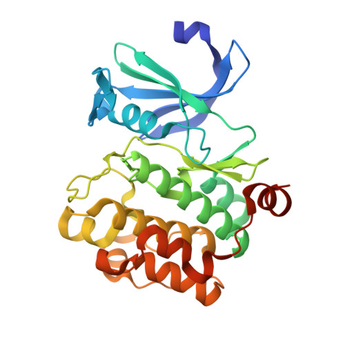
4MTA

Crystal structure of Pim-1 kinase domain in complex with 2-methyl-5-phenylfuran-3-carboxylic acid


Experimental Data Snapshot

  • Method: X-RAY DIFFRACTION
  • Resolution: 2.20 Å
  • R-Value Free: 0.192 
  • R-Value Work: 0.158 
  • R-Value Observed: 0.160 

wwPDB Validation   3D Report Full Report


Ligand Structure Quality Assessment 


This is version 1.2 of the entry. See complete history


Literature

Large scale meta-analysis of fragment-based screening campaigns: privileged fragments and complementary technologies.

Kutchukian, P.S.Wassermann, A.M.Lindvall, M.K.Wright, S.K.Ottl, J.Jacob, J.Scheufler, C.Marzinzik, A.Brooijmans, N.Glick, M.

(2015) J Biomol Screen 20: 588-596

  • DOI: https://doi.org/10.1177/1087057114565080
  • Primary Citation of Related Structures:  
    4MTA

  • PubMed Abstract: 

    A first step in fragment-based drug discovery (FBDD) often entails a fragment-based screen (FBS) to identify fragment "hits." However, the integration of conflicting results from orthogonal screens remains a challenge. Here we present a meta-analysis of 35 fragment-based campaigns at Novartis, which employed a generic 1400-fragment library against diverse target families using various biophysical and biochemical techniques. By statistically interrogating the multidimensional FBS data, we sought to investigate three questions: (1) What makes a fragment amenable for FBS? (2) How do hits from different fragment screening technologies and target classes compare with each other? (3) What is the best way to pair FBS assay technologies? In doing so, we identified substructures that were privileged for specific target classes, as well as fragments that were privileged for authentic activity against many targets. We also revealed some of the discrepancies between technologies. Finally, we uncovered a simple rule of thumb in screening strategy: when choosing two technologies for a campaign, pairing a biochemical and biophysical screen tends to yield the greatest coverage of authentic hits.


  • Organizational Affiliation

    Novartis Institutes for BioMedical Research, Cambridge, MA, USA Current address: Merck, Boston, MA, USA.


Macromolecules
Find similar proteins by:  (by identity cutoff)  |  3D Structure
Entity ID: 1
MoleculeChains Sequence LengthOrganismDetailsImage
Serine/threonine-protein kinase pim-1286Homo sapiensMutation(s): 0 
Gene Names: PIM1
EC: 2.7.11.1
UniProt & NIH Common Fund Data Resources
Find proteins for P11309 (Homo sapiens)
Explore P11309 
Go to UniProtKB:  P11309
PHAROS:  P11309
GTEx:  ENSG00000137193 
Entity Groups  
Sequence Clusters30% Identity50% Identity70% Identity90% Identity95% Identity100% Identity
UniProt GroupP11309
Sequence Annotations
Expand
  • Reference Sequence
Small Molecules
Ligands 2 Unique
IDChains Name / Formula / InChI Key2D Diagram3D Interactions
2D2
Query on 2D2

Download Ideal Coordinates CCD File 
B [auth A]2-methyl-5-phenylfuran-3-carboxylic acid
C12 H10 O3
VLMNACSEESRUAK-UHFFFAOYSA-N
GOL
Query on GOL

Download Ideal Coordinates CCD File 
C [auth A]GLYCEROL
C3 H8 O3
PEDCQBHIVMGVHV-UHFFFAOYSA-N
Experimental Data & Validation

Experimental Data

  • Method: X-RAY DIFFRACTION
  • Resolution: 2.20 Å
  • R-Value Free: 0.192 
  • R-Value Work: 0.158 
  • R-Value Observed: 0.160 
  • Space Group: P 65
Unit Cell:
Length ( Å )Angle ( ˚ )
a = 95.79α = 90
b = 95.79β = 90
c = 79.76γ = 120
Software Package:
Software NamePurpose
PHASERphasing
BUSTERrefinement
XDSdata reduction
XSCALEdata scaling

Structure Validation

View Full Validation Report


Ligand Structure Quality Assessment 


Entry History 

Deposition Data

Revision History  (Full details and data files)

  • Version 1.0: 2015-01-14
    Type: Initial release
  • Version 1.1: 2015-06-03
    Changes: Database references
  • Version 1.2: 2023-09-20
    Changes: Data collection, Database references, Derived calculations, Refinement description