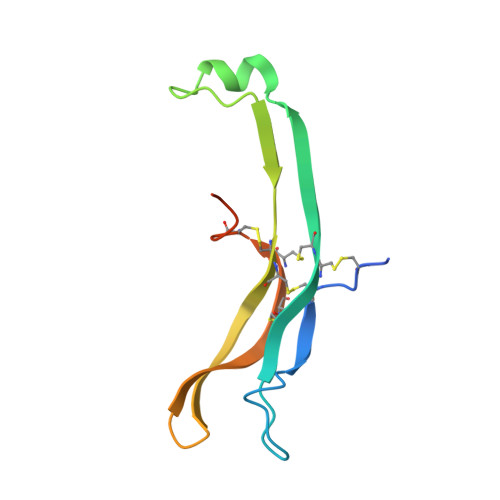
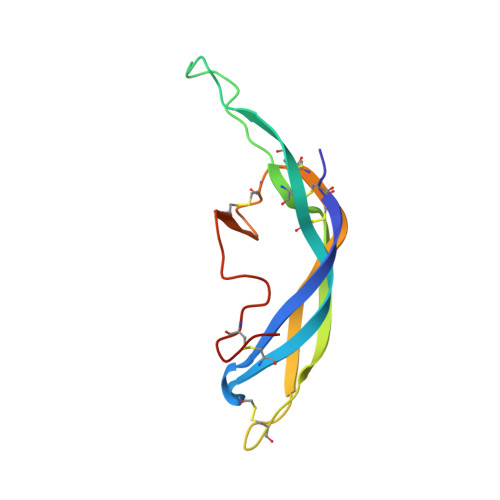
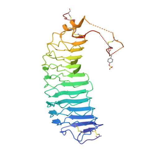
4MQW

Structure of follicle-stimulating hormone in complex with the entire ectodomain of its receptor (P31)


Experimental Data Snapshot

  • Method: X-RAY DIFFRACTION
  • Resolution: 2.90 Å
  • R-Value Free: 0.237 
  • R-Value Work: 0.174 
  • R-Value Observed: 0.177 

wwPDB Validation   3D Report Full Report


Ligand Structure Quality Assessment 


This is version 1.6 of the entry. See complete history


Literature

Evidence for Follicle-stimulating Hormone Receptor as a Functional Trimer.

Jiang, X.Fischer, D.Chen, X.McKenna, S.D.Liu, H.Sriraman, V.Yu, H.N.Goutopoulos, A.Arkinstall, S.He, X.

(2014) J Biol Chem 289: 14273-14282

  • DOI: https://doi.org/10.1074/jbc.M114.549592
  • Primary Citation of Related Structures:  
    4MQW

  • PubMed Abstract: 

    Follicle-stimulating hormone receptor (FSHR), a G-protein coupled receptor, is an important drug target in the development of novel therapeutics for reproductive indications. The FSHR extracellular domains were observed in the crystal structure as a trimer, which enabled us to propose a novel model for the receptor activation mechanism. The model predicts that FSHR binds Asnα(52)-deglycosylated FSH at a 3-fold higher capacity than fully glycosylated FSH. It also predicts that, upon dissociation of the FSHR trimer into monomers, the binding of glycosylated FSH, but not deglycosylated FSH, would increase 3-fold, and that the dissociated monomers would in turn enhance FSHR binding and signaling activities by 3-fold. This study presents evidence confirming these predictions and provides crystallographic and mutagenesis data supporting the proposed model. The model also provides a mechanistic explanation to the agonist and antagonist activities of thyroid-stimulating hormone receptor autoantibodies. We conclude that FSHR exists as a functional trimer.


  • Organizational Affiliation

    From the EMD Serono Research and Development Institute, Billerica, Massachusetts 01821 and xuliang.jiang@emdserono.com.


Macromolecules
Find similar proteins by:  (by identity cutoff)  |  3D Structure
Entity ID: 1
MoleculeChains Sequence LengthOrganismDetailsImage
Glycoprotein hormones, alpha polypeptide
A, D, G
102Homo sapiensMutation(s): 0 
Gene Names: CGA
UniProt & NIH Common Fund Data Resources
Find proteins for P01215 (Homo sapiens)
Explore P01215 
Go to UniProtKB:  P01215
PHAROS:  P01215
GTEx:  ENSG00000135346 
Entity Groups  
Sequence Clusters30% Identity50% Identity70% Identity90% Identity95% Identity100% Identity
UniProt GroupP01215
Sequence Annotations
Expand
  • Reference Sequence
Find similar proteins by:  (by identity cutoff)  |  3D Structure
Entity ID: 2
MoleculeChains Sequence LengthOrganismDetailsImage
Follitropin subunit beta
B, E, H
111Homo sapiensMutation(s): 0 
Gene Names: FSHB
UniProt & NIH Common Fund Data Resources
Find proteins for P01225 (Homo sapiens)
Explore P01225 
Go to UniProtKB:  P01225
PHAROS:  P01225
GTEx:  ENSG00000131808 
Entity Groups  
Sequence Clusters30% Identity50% Identity70% Identity90% Identity95% Identity100% Identity
UniProt GroupP01225
Sequence Annotations
Expand
  • Reference Sequence
Find similar proteins by:  (by identity cutoff)  |  3D Structure
Entity ID: 3
MoleculeChains Sequence LengthOrganismDetailsImage
Follicle-stimulating hormone receptorC [auth X],
F [auth Y],
I [auth Z]
361Homo sapiensMutation(s): 1 
Gene Names: FSHRLGR1
UniProt & NIH Common Fund Data Resources
Find proteins for P23945 (Homo sapiens)
Explore P23945 
Go to UniProtKB:  P23945
PHAROS:  P23945
GTEx:  ENSG00000170820 
Entity Groups  
Sequence Clusters30% Identity50% Identity70% Identity90% Identity95% Identity100% Identity
UniProt GroupP23945
Sequence Annotations
Expand
  • Reference Sequence
Small Molecules
Ligands 3 Unique
IDChains Name / Formula / InChI Key2D Diagram3D Interactions
JEF
Query on JEF

Download Ideal Coordinates CCD File 
CA [auth Z]O-(O-(2-AMINOPROPYL)-O'-(2-METHOXYETHYL)POLYPROPYLENE GLYCOL 500)
C30 H63 N O10
ICCXIDTYQFYPNV-RUMGZKRTSA-N
NAG
Query on NAG

Download Ideal Coordinates CCD File 
AA [auth H]
BA [auth H]
DA [auth Z]
J [auth A]
K [auth A]
AA [auth H],
BA [auth H],
DA [auth Z],
J [auth A],
K [auth A],
L [auth B],
M [auth B],
R [auth X],
S [auth D],
T [auth D],
U [auth E],
V [auth E],
X [auth Y],
Y [auth G],
Z [auth G]
2-acetamido-2-deoxy-beta-D-glucopyranose
C8 H15 N O6
OVRNDRQMDRJTHS-FMDGEEDCSA-N
EDO
Query on EDO

Download Ideal Coordinates CCD File 
N [auth B],
O [auth B],
P [auth X],
Q [auth X],
W [auth Y]
1,2-ETHANEDIOL
C2 H6 O2
LYCAIKOWRPUZTN-UHFFFAOYSA-N
Modified Residues  1 Unique
IDChains TypeFormula2D DiagramParent
TYS
Query on TYS
C [auth X],
F [auth Y],
I [auth Z]
L-PEPTIDE LINKINGC9 H11 N O6 STYR
Experimental Data & Validation

Experimental Data

  • Method: X-RAY DIFFRACTION
  • Resolution: 2.90 Å
  • R-Value Free: 0.237 
  • R-Value Work: 0.174 
  • R-Value Observed: 0.177 
  • Space Group: P 31
Unit Cell:
Length ( Å )Angle ( ˚ )
a = 95.9α = 90
b = 95.9β = 90
c = 204.281γ = 120
Software Package:
Software NamePurpose
HKL-3000data collection
PHASESphasing
REFMACrefinement
HKL-3000data reduction
HKL-3000data scaling
BUSTERrefinement

Structure Validation

View Full Validation Report


Ligand Structure Quality Assessment 


Entry History 

Deposition Data

Revision History  (Full details and data files)

  • Version 1.0: 2014-04-09
    Type: Initial release
  • Version 1.1: 2014-06-18
    Changes: Database references
  • Version 1.2: 2014-09-24
    Changes: Database references
  • Version 1.3: 2020-07-29
    Type: Remediation
    Reason: Carbohydrate remediation
    Changes: Data collection, Database references, Derived calculations, Structure summary
  • Version 1.4: 2023-09-20
    Changes: Data collection, Database references, Refinement description, Structure summary
  • Version 1.5: 2023-12-06
    Changes: Data collection
  • Version 1.6: 2024-01-10
    Changes: Derived calculations