


4MPK
Crystal structure of the complex of buffalo signaling protein SPB-40 with N-acetylglucosamine at 2.65 A resolution
- PDB DOI: https://doi.org/10.2210/pdb4MPK/pdb
- Classification: SIGNALING PROTEIN
- Organism(s): Bubalus bubalis
- Mutation(s): No
- Deposited: 2013-09-13 Released: 2013-11-06
Experimental Data Snapshot
- Method: X-RAY DIFFRACTION
- Resolution: 2.65 Å
- R-Value Free: 0.234
- R-Value Work: 0.174
- R-Value Observed: 0.177

This is version 1.2 of the entry. See complete history.

Macromolecules
Find similar proteins by:
(by identity cutoff) | 3D Structure
Entity ID: 1 | |||||
|---|---|---|---|---|---|
| Molecule | Chains | Sequence Length | Organism | Details | Image |
| Chitinase-3-like protein 1 | 361 | Bubalus bubalis | Mutation(s): 0 | 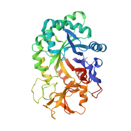 | |
UniProt | |||||
Find proteins for Q7YS85 (Bubalus bubalis) Explore Q7YS85 Go to UniProtKB: Q7YS85 | |||||
Entity Groups | |||||
| Sequence Clusters | 30% Identity50% Identity70% Identity90% Identity95% Identity100% Identity | ||||
| UniProt Group | Q7YS85 | ||||
Sequence AnnotationsExpand | |||||
| |||||
Small Molecules
| Ligands 2 Unique | |||||
|---|---|---|---|---|---|
| ID | Chains | Name / Formula / InChI Key | 2D Diagram | 3D Interactions | |
| NAG Query on NAG | B [auth A], C [auth A] | 2-acetamido-2-deoxy-beta-D-glucopyranose C8 H15 N O6 OVRNDRQMDRJTHS-FMDGEEDCSA-N |  | ||
| GOL Query on GOL | D [auth A] | GLYCEROL C3 H8 O3 PEDCQBHIVMGVHV-UHFFFAOYSA-N |  | ||
Experimental Data & Validation
Experimental Data
- Method: X-RAY DIFFRACTION
- Resolution: 2.65 Å
- R-Value Free: 0.234
- R-Value Work: 0.174
- R-Value Observed: 0.177
- Space Group: P 21 21 21
Unit Cell:
| Length ( Å ) | Angle ( ˚ ) |
|---|---|
| a = 60.938 | α = 90 |
| b = 67.11 | β = 90 |
| c = 107.006 | γ = 90 |
| Software Name | Purpose |
|---|---|
| HKL-2000 | data collection |
| AMoRE | phasing |
| REFMAC | refinement |
| DENZO | data reduction |
| SCALEPACK | data scaling |
Entry History
Deposition Data
- Released Date: 2013-11-06 Deposition Author(s): Yamini, S., Chaudhary, A., Sinha, M., Kaur, P., Sharma, S., Singh, T.P.
Revision History (Full details and data files)
- Version 1.0: 2013-11-06
Type: Initial release - Version 1.1: 2020-07-29
Type: Remediation
Reason: Carbohydrate remediation
Changes: Data collection, Derived calculations, Structure summary - Version 1.2: 2023-11-08
Changes: Data collection, Database references, Refinement description, Structure summary
