


Structure of Zingiber officinale double bond reductase
Buratto, J., Langlois d'Estaintot, B., Granier, T., Gallois, B., Willis, M.A., Sang, Y., Flores-Sanchez, I.J., Gang, D.R.To be published.
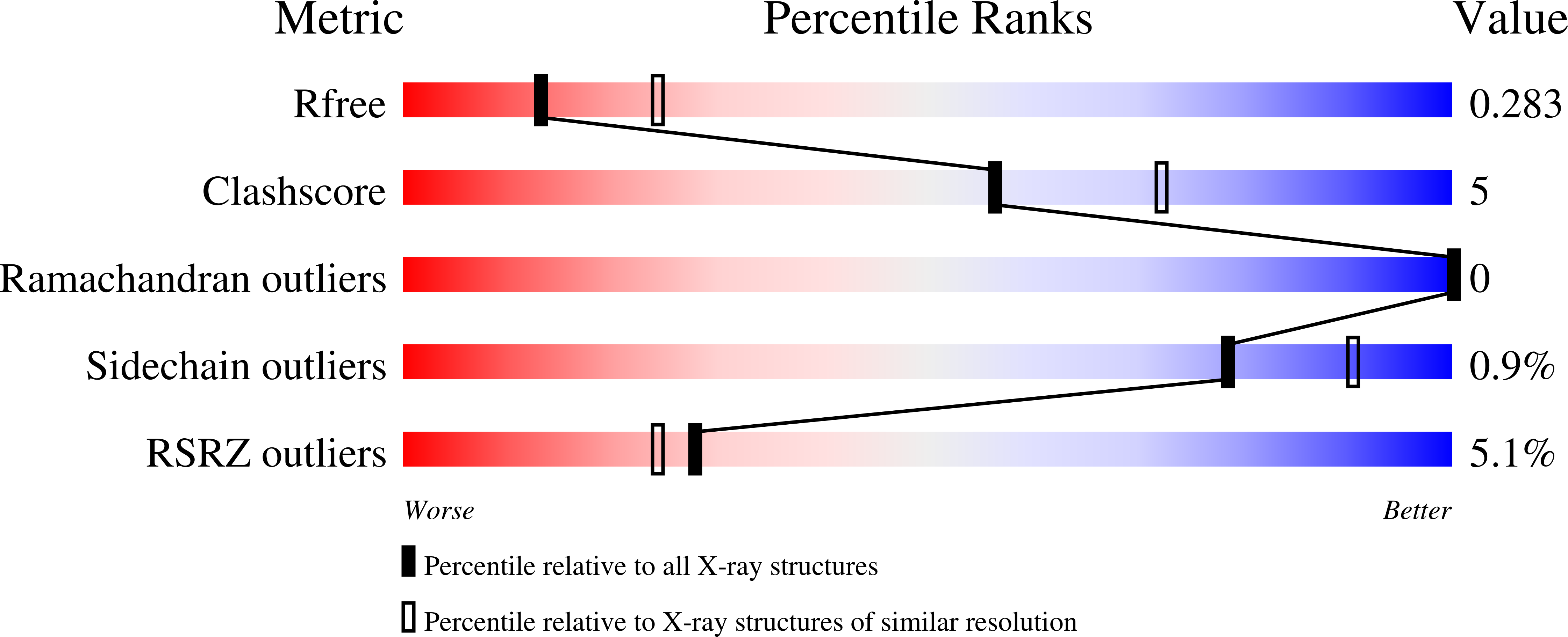
Experimental Data Snapshot
wwPDB Validation 3D Report Full Report
Entity ID: 1 | |||||
|---|---|---|---|---|---|
| Molecule | Chains | Sequence Length | Organism | Details | Image |
| Zingiber officinale double bond reductase | 358 | Zingiber officinale | Mutation(s): 0 | 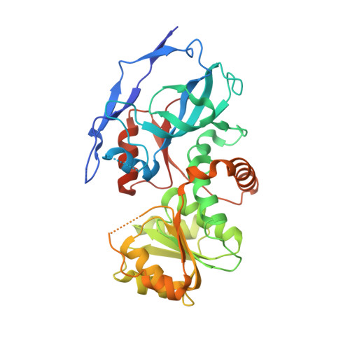 | |
UniProt | |||||
Find proteins for A0A096LNF0 (Zingiber officinale) Explore A0A096LNF0 Go to UniProtKB: A0A096LNF0 | |||||
Entity Groups | |||||
| Sequence Clusters | 30% Identity50% Identity70% Identity90% Identity95% Identity100% Identity | ||||
| UniProt Group | A0A096LNF0 | ||||
Sequence AnnotationsExpand | |||||
| |||||
| Length ( Å ) | Angle ( ˚ ) |
|---|---|
| a = 50.93 | α = 80.15 |
| b = 76.34 | β = 89.97 |
| c = 93.45 | γ = 85.66 |
| Software Name | Purpose |
|---|---|
| XSCALE | data scaling |
| PHASER | phasing |
| REFMAC | refinement |
| PDB_EXTRACT | data extraction |
| MxCuBE | data collection |
| XDS | data reduction |
RCSB PDB (citation) is hosted by
RCSB PDB is a member of the

