4MJO

Human liver fructose-1,6-bisphosphatase(d-fructose-1,6-bisphosphate, 1-phosphohydrolase) (e.c.3.1.3.11) complexed with the allosteric inhibitor 3


Experimental Data Snapshot

  • Method: X-RAY DIFFRACTION
  • Resolution: 2.40 Å
  • R-Value Free: 0.236 
  • R-Value Work: 0.207 
  • R-Value Observed: 0.208 

wwPDB Validation   3D Report Full Report


Ligand Structure Quality Assessment 


This is version 1.3 of the entry. See complete history


Literature

Determination of protein-ligand binding constants of a cooperatively regulated tetrameric enzyme using electrospray mass spectrometry.

Cubrilovic, D.Haap, W.Barylyuk, K.Ruf, A.Badertscher, M.Gubler, M.Tetaz, T.Joseph, C.Benz, J.Zenobi, R.

(2014) ACS Chem Biol 9: 218-226

  • DOI: https://doi.org/10.1021/cb4007002
  • Primary Citation of Related Structures:  
    4MJO

  • PubMed Abstract: 

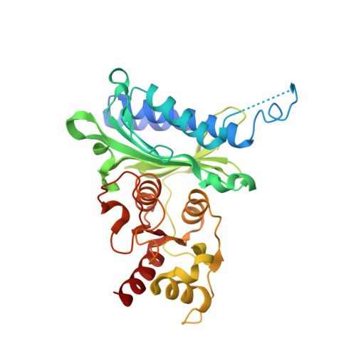
    This study highlights the benefits of nano electrospray ionization mass spectrometry (nanoESI-MS) as a fast and label-free method not only for determination of dissociation constants (KD) of a cooperatively regulated enzyme but also to better understand the mechanism of enzymatic cooperativity of multimeric proteins. We present an approach to investigate the allosteric mechanism in the binding of inhibitors to the homotetrameric enzyme fructose 1,6-bisphosphatase (FBPase), a potential therapeutic target for glucose control in type 2 diabetes. A series of inhibitors binding at an allosteric site of FBPase were investigated to determine their KDs by nanoESI-MS. The KDs determined by ESI-MS correlate very well with IC50 values in solution. The Hill coefficients derived from nanoESI-MS suggest positive cooperativity. From single-point measurements we could obtain information on relative potency, stoichiometry, conformational changes, and mechanism of cooperativity. A new X-ray crystal structure of FBPase tetramer binding ligand 3 in a 4:4 stoichiometry is also reported. NanoESI-MS-based results match the current understanding of the investigated system and are in agreement with the X-ray structural data, but provide additional mechanistic insight on the ligand binding, due to the better dynamic resolution. This method offers a powerful approach for studying other proteins with allosteric binding sites, as well.


  • Organizational Affiliation

    Department of Chemistry and Applied Biosciences, ETH Zurich , 8093 Zurich, Switzerland.


Macromolecules
Find similar proteins by:  (by identity cutoff)  |  3D Structure
Entity ID: 1
MoleculeChains Sequence LengthOrganismDetailsImage
Fructose-1,6-bisphosphatase 1
A, B, C, D, E
A, B, C, D, E, F, G, H
338Homo sapiensMutation(s): 0 
EC: 3.1.3.11
UniProt & NIH Common Fund Data Resources
Find proteins for P09467 (Homo sapiens)
Explore P09467 
Go to UniProtKB:  P09467
PHAROS:  P09467
GTEx:  ENSG00000165140 
Entity Groups  
Sequence Clusters30% Identity50% Identity70% Identity90% Identity95% Identity100% Identity
UniProt GroupP09467
Sequence Annotations
Expand
  • Reference Sequence
Small Molecules
Binding Affinity Annotations 
IDSourceBinding Affinity
2C1 PDBBind:  4MJO Kd: 600 (nM) from 1 assay(s)
Binding MOAD:  4MJO Kd: 600 (nM) from 1 assay(s)
BindingDB:  4MJO IC50: min: 80, max: 1000 (nM) from 2 assay(s)
Experimental Data & Validation

Experimental Data

  • Method: X-RAY DIFFRACTION
  • Resolution: 2.40 Å
  • R-Value Free: 0.236 
  • R-Value Work: 0.207 
  • R-Value Observed: 0.208 
  • Space Group: P 1 21 1
Unit Cell:
Length ( Å )Angle ( ˚ )
a = 65.974α = 90
b = 286.305β = 97.79
c = 83.692γ = 90
Software Package:
Software NamePurpose
REFMACrefinement

Structure Validation

View Full Validation Report


Ligand Structure Quality Assessment 


Entry History 

Deposition Data

Revision History  (Full details and data files)

  • Version 1.0: 2013-11-06
    Type: Initial release
  • Version 1.1: 2014-01-29
    Changes: Database references
  • Version 1.2: 2018-04-25
    Changes: Advisory, Data collection, Source and taxonomy
  • Version 1.3: 2023-09-20
    Changes: Data collection, Database references, Derived calculations, Refinement description