Signature motifs identify an acinetobacter cif virulence factor with epoxide hydrolase activity.
Bahl, C.D., Hvorecny, K.L., Bridges, A.A., Ballok, A.E., Bomberger, J.M., Cady, K.C., O'Toole, G.A., Madden, D.R.(2014) J Biol Chem 289: 7460-7469
- PubMed: 24474692
- DOI: https://doi.org/10.1074/jbc.M113.518092
- Primary Citation of Related Structures:
4MEA, 4MEB - PubMed Abstract:
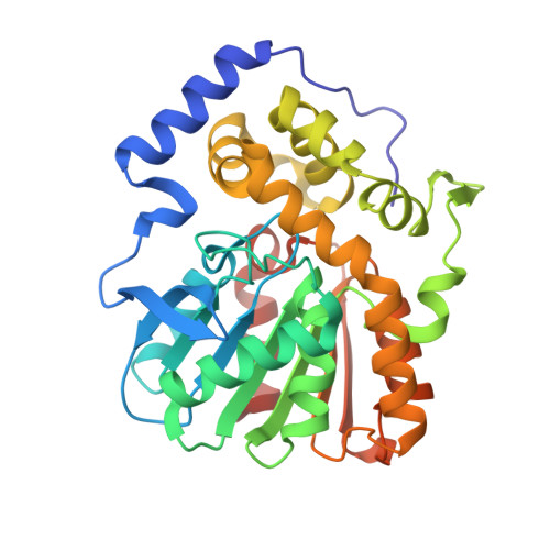
Endocytic recycling of the cystic fibrosis transmembrane conductance regulator (CFTR) is blocked by the CFTR inhibitory factor (Cif). Originally discovered in Pseudomonas aeruginosa, Cif is a secreted epoxide hydrolase that is transcriptionally regulated by CifR, an epoxide-sensitive repressor. In this report, we investigate a homologous protein found in strains of the emerging nosocomial pathogens Acinetobacter nosocomialis and Acinetobacter baumannii ("aCif"). Like Cif, aCif is an epoxide hydrolase that carries an N-terminal secretion signal and can be purified from culture supernatants. When applied directly to polarized airway epithelial cells, mature aCif triggers a reduction in CFTR abundance at the apical membrane. Biochemical and crystallographic studies reveal a dimeric assembly with a stereochemically conserved active site, confirming our motif-based identification of candidate Cif-like pathogenic EH sequences. Furthermore, cif expression is transcriptionally repressed by a CifR homolog ("aCifR") and is induced in the presence of epoxides. Overall, this Acinetobacter protein recapitulates the essential attributes of the Pseudomonas Cif system and thus may facilitate airway colonization in nosocomial lung infections.
Organizational Affiliation:
From the Departments of Biochemistry and.