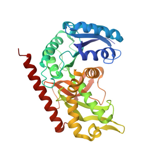
Refined crystal structure of cytoplasmic malate dehydrogenase at 2.5-A resolution.
Birktoft, J.J., Rhodes, G., Banaszak, L.J.(1989) Biochemistry 28: 6065-6081
- PubMed: 2775751
- DOI: https://doi.org/10.1021/bi00440a051
- Primary Citation of Related Structures:
4MDH - PubMed Abstract:
The molecular structure of cytoplasmic malate dehydrogenase from pig heart has been refined by alternating rounds of restrained least-squares methods and model readjustment on an interactive graphics system. The resulting structure contains 333 amino acids in each of the two subunits, 2 NAD molecules, 471 solvent molecules, and 2 large noncovalently bound molecules that are assumed to be sulfate ions. The crystallographic study was done on one entire dimer without symmetry restraints. Analysis of the relative position of the two subunits shows that the dimer does not obey exact 2-fold rotational symmetry; instead, the subunits are related by a 173 degrees rotation. The structure results in a R factor of 16.7% for diffraction data between 6.0 and 2.5 A, and the rms deviations from ideal bond lengths and angles are 0.017 A and 2.57 degrees, respectively. The bound coenzyme in addition to hydrophobic interactions makes numerous hydrogen bonds that either are directly between NAD and the enzyme or are with solvent molecules, some of which in turn are hydrogen bonded to the enzyme. The carboxamide group of NAD is hydrogen bonded to the side chain of Asn-130 and via a water molecule to the backbone nitrogens of Leu-157 and Asp-158 and to the carbonyl oxygen of Leu-154. Asn-130 is one of the corner residues in a beta-turn that contains the lone cis peptide bond in cytoplasmic malate dehydrogenase, situated between Asn-130 and Pro-131. The active site histidine, His-186, is hydrogen bonded from nitrogen ND1 to the carboxylate of Asp-158 and from its nitrogen NE2 to the sulfate ion bound in the putative substrate binding site. In addition to interacting with the active site histidine, this sulfate ion is also hydrogen bonded to the guanidinium group of Arg-161, to the carboxamide group of Asn-140, and to the hydroxyl group of Ser-241. It is speculated that the substrate, malate or oxaloacetate, is bound in the sulfate binding site with the substrate 1-carboxyl hydrogen bonded to the guanidinium group of Arg-161.
Organizational Affiliation:
Department of Biological Chemistry, Washington University School of Medicine, St. Louis, Missouri 63110.