4MCW

Crystal structure of a HD-GYP domain (a cyclic-di-GMP phosphodiesterase) containing a tri-nuclear metal centre


Experimental Data Snapshot

  • Method: X-RAY DIFFRACTION
  • Resolution: 2.03 Å
  • R-Value Free: 0.213 
  • R-Value Work: 0.187 
  • R-Value Observed: 0.189 

wwPDB Validation   3D Report Full Report


This is version 1.2 of the entry. See complete history


Literature

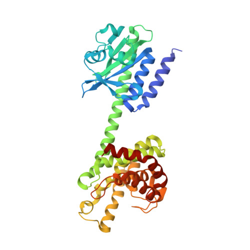
Crystal structure of an HD-GYP domain cyclic-di-GMP phosphodiesterase reveals an enzyme with a novel trinuclear catalytic iron centre.

Bellini, D.Caly, D.L.McCarthy, Y.Bumann, M.An, S.Q.Dow, J.M.Ryan, R.P.Walsh, M.A.

(2014) Mol Microbiol 91: 26-38

  • DOI: https://doi.org/10.1111/mmi.12447
  • Primary Citation of Related Structures:  
    4MCW, 4MDZ, 4ME4

  • PubMed Abstract: 

    Bis-(3',5') cyclic di-guanylate (c-di-GMP) is a key bacterial second messenger that is implicated in the regulation of many crucial processes that include biofilm formation, motility and virulence. Cellular levels of c-di-GMP are controlled through synthesis by GGDEF domain diguanylate cyclases and degradation by two classes of phosphodiesterase with EAL or HD-GYP domains. Here, we have determined the structure of an enzymatically active HD-GYP domain protein from Persephonella marina (PmGH) alone, in complex with substrate (c-di-GMP) and final reaction product (GMP). The structures reveal a novel trinuclear iron binding site, which is implicated in catalysis and identify residues involved in recognition of c-di-GMP. This structure completes the picture of all domains involved in c-di-GMP metabolism and reveals that the HD-GYP family splits into two distinct subgroups containing bi- and trinuclear metal centres.


  • Organizational Affiliation

    Diamond Light Source, Harwell Science and Innovation Campus, Didcot, Oxfordshire, OX11 0DE, UK; Research Complex at Harwell, Harwell Science and Innovation Campus, Didcot, Oxfordshire, OX11 0FA, UK.


Macromolecules
Find similar proteins by:  (by identity cutoff)  |  3D Structure
Entity ID: 1
MoleculeChains Sequence LengthOrganismDetailsImage
Metal dependent phosphohydrolase
A, B
369Persephonella marina EX-H1Mutation(s): 2 
Gene Names: PERMA_0986
EC: 3.1.4.1
UniProt
Find proteins for C0QQ26 (Persephonella marina (strain DSM 14350 / EX-H1))
Explore C0QQ26 
Go to UniProtKB:  C0QQ26
Entity Groups  
Sequence Clusters30% Identity50% Identity70% Identity90% Identity95% Identity100% Identity
UniProt GroupC0QQ26
Sequence Annotations
Expand
  • Reference Sequence
Small Molecules
Ligands 4 Unique
IDChains Name / Formula / InChI Key2D Diagram3D Interactions
SIN
Query on SIN

Download Ideal Coordinates CCD File 
F [auth A]
G [auth A]
H [auth A]
N [auth B]
O [auth B]
F [auth A],
G [auth A],
H [auth A],
N [auth B],
O [auth B],
P [auth B]
SUCCINIC ACID
C4 H6 O4
KDYFGRWQOYBRFD-UHFFFAOYSA-N
IMD
Query on IMD

Download Ideal Coordinates CCD File 
I [auth A]IMIDAZOLE
C3 H5 N2
RAXXELZNTBOGNW-UHFFFAOYSA-O
EDO
Query on EDO

Download Ideal Coordinates CCD File 
J [auth A]1,2-ETHANEDIOL
C2 H6 O2
LYCAIKOWRPUZTN-UHFFFAOYSA-N
FE
Query on FE

Download Ideal Coordinates CCD File 
C [auth A]
D [auth A]
E [auth A]
K [auth B]
L [auth B]
C [auth A],
D [auth A],
E [auth A],
K [auth B],
L [auth B],
M [auth B]
FE (III) ION
Fe
VTLYFUHAOXGGBS-UHFFFAOYSA-N
Experimental Data & Validation

Experimental Data

  • Method: X-RAY DIFFRACTION
  • Resolution: 2.03 Å
  • R-Value Free: 0.213 
  • R-Value Work: 0.187 
  • R-Value Observed: 0.189 
  • Space Group: I 2 2 2
Unit Cell:
Length ( Å )Angle ( ˚ )
a = 69.83α = 90
b = 182.38β = 90
c = 232.47γ = 90
Software Package:
Software NamePurpose
EDNAdata collection
SHELXmodel building
REFMACrefinement
XDSdata reduction
Aimlessdata scaling
SHELXphasing

Structure Validation

View Full Validation Report


Entry History 

Deposition Data

Revision History  (Full details and data files)

  • Version 1.0: 2014-02-19
    Type: Initial release
  • Version 1.1: 2021-08-11
    Changes: Database references, Derived calculations, Structure summary
  • Version 1.2: 2024-02-28
    Changes: Data collection