4M9W

Crystal Structure of Ara h 8 with MES bound


Experimental Data Snapshot

  • Method: X-RAY DIFFRACTION
  • Resolution: 1.95 Å
  • R-Value Free: 0.248 
  • R-Value Work: 0.205 
  • R-Value Observed: 0.208 

wwPDB Validation   3D Report Full Report


This is version 1.2 of the entry. See complete history


Literature

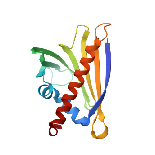
Structure and Function of the Peanut Panallergen Ara h 8.

Hurlburt, B.K.Offermann, L.R.McBride, J.K.Majorek, K.A.Maleki, S.J.Chruszcz, M.

(2013) J Biol Chem 288: 36890-36901

  • DOI: https://doi.org/10.1074/jbc.M113.517797
  • Primary Citation of Related Structures:  
    4M9B, 4M9W, 4MA6, 4MAP

  • PubMed Abstract: 

    The incidence of peanut allergy continues to rise in the United States and Europe. Whereas exposure to the major allergens Ara h 1, 2, 3, and 6 can cause fatal anaphylaxis, exposure to the minor allergens usually does not. Ara h 8 is a minor allergen. Importantly, it is the minor food allergens that are thought to be responsible for oral allergy syndrome (OAS), in which sensitization to airborne allergens causes a Type 2 allergic reaction to ingested foods. Furthermore, it is believed that similar protein structure rather than a similar linear sequence is the cause of OAS. Bet v 1 from birch pollen is a common sensitizing agent, and OAS results when patients consume certain fruits, vegetables, tree nuts, and peanuts. Here, we report the three-dimensional structure of Ara h 8, a Bet v 1 homolog. The overall fold is very similar to that of Bet v 1, Api g 1 (celery), Gly m 4 (soy), and Pru av 1 (cherry). Ara h 8 binds the isoflavones quercetin and apigenin as well as resveratrol avidly.


  • Organizational Affiliation

    From the Southern Regional Research Center, Agricultural Research Service, United States Department of Agriculture, New Orleans, Louisiana 70124.


Macromolecules
Find similar proteins by:  (by identity cutoff)  |  3D Structure
Entity ID: 1
MoleculeChains Sequence LengthOrganismDetailsImage
Ara h 8 allergen
A, B
157Arachis hypogaeaMutation(s): 0 
UniProt
Find proteins for Q6VT83 (Arachis hypogaea)
Explore Q6VT83 
Go to UniProtKB:  Q6VT83
Entity Groups  
Sequence Clusters30% Identity50% Identity70% Identity90% Identity95% Identity100% Identity
UniProt GroupQ6VT83
Sequence Annotations
Expand
  • Reference Sequence
Experimental Data & Validation

Experimental Data

  • Method: X-RAY DIFFRACTION
  • Resolution: 1.95 Å
  • R-Value Free: 0.248 
  • R-Value Work: 0.205 
  • R-Value Observed: 0.208 
  • Space Group: P 1 21 1
Unit Cell:
Length ( Å )Angle ( ˚ )
a = 40.183α = 90
b = 87.488β = 95.84
c = 42.29γ = 90
Software Package:
Software NamePurpose
HKL-3000data collection
HKL-3000phasing
MOLREPphasing
REFMACrefinement
HKL-2000data reduction
HKL-2000data scaling

Structure Validation

View Full Validation Report


Entry History 

Deposition Data

Revision History  (Full details and data files)

  • Version 1.0: 2013-11-27
    Type: Initial release
  • Version 1.1: 2014-01-15
    Changes: Database references
  • Version 1.2: 2023-09-20
    Changes: Data collection, Database references, Derived calculations, Refinement description