4M6F

Dimer of the G-Segment Invertase bound to a DNA substrate


Experimental Data Snapshot

  • Method: X-RAY DIFFRACTION
  • Resolution: 4.99 Å
  • R-Value Free: 0.288 
  • R-Value Work: 0.229 
  • R-Value Observed: 0.230 

wwPDB Validation   3D Report Full Report


This is version 1.3 of the entry. See complete history


Literature

Exploiting large non-isomorphous differences for phase determination of a G-segment invertase-DNA complex.

Ritacco, C.J.Steitz, T.A.Wang, J.

(2014) Acta Crystallogr D Biol Crystallogr 70: 685-693

  • DOI: https://doi.org/10.1107/S1399004713032392
  • Primary Citation of Related Structures:  
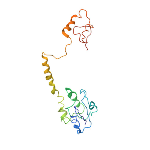
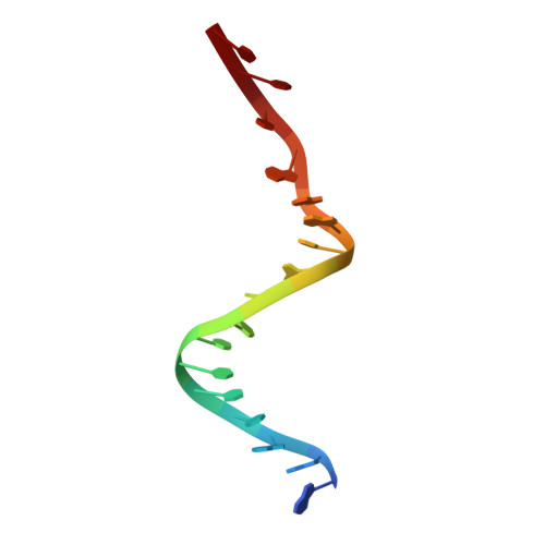
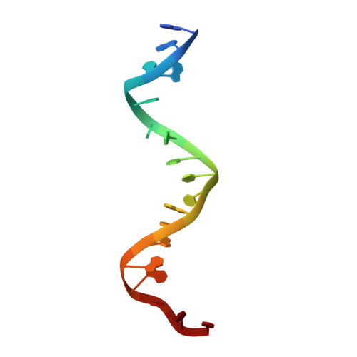
    4M6F

  • PubMed Abstract: 

    Crystals of the G-segment invertase in complex with a 37-base-pair asymmetric DNA duplex substrate had an unusually high solvent content of 88% and diffracted to a maximal resolution of about 5.0 Å. These crystals exhibited a high degree of non-isomorphism and anisotropy, which presented a serious challenge for structure determination by isomorphous replacement. Here, a procedure of cross-crystal averaging is described that uses large non-isomorphous crystallographic data with a priori information of an approximate molecular boundary as determined from a minimal amount of experimental phase information. Using this procedure, high-quality experimental phases were obtained that have enabled it to be shown that the conformation of the bound substrate DNA duplex significantly differs from those of substrates bound in other serine recombinase-DNA complexes.


  • Organizational Affiliation

    Department of Molecular Biophysics and Biochemistry, Yale University, New Haven, CT 06520, USA.


Macromolecules

Find similar proteins by:  (by identity cutoff)  |  3D Structure
Entity ID: 1
MoleculeChains Sequence LengthOrganismDetailsImage
DNA-invertase193Muvirus muMutation(s): 0 
UniProt
Find proteins for P03015 (Escherichia phage Mu)
Explore P03015 
Go to UniProtKB:  P03015
Entity Groups  
Sequence Clusters30% Identity50% Identity70% Identity90% Identity95% Identity100% Identity
UniProt GroupP03015
Sequence Annotations
Expand
  • Reference Sequence

Find similar nucleic acids by:  Sequence   |   3D Structure  

Entity ID: 2
MoleculeChains LengthOrganismImage
gix site analog17synthetic construct
Sequence Annotations
Expand
  • Reference Sequence

Find similar nucleic acids by:  Sequence   |   3D Structure  

Entity ID: 3
MoleculeChains LengthOrganismImage
gix site analog18synthetic construct
Sequence Annotations
Expand
  • Reference Sequence
Experimental Data & Validation

Experimental Data

  • Method: X-RAY DIFFRACTION
  • Resolution: 4.99 Å
  • R-Value Free: 0.288 
  • R-Value Work: 0.229 
  • R-Value Observed: 0.230 
  • Space Group: P 64 2 2
Unit Cell:
Length ( Å )Angle ( ˚ )
a = 119.8α = 90
b = 119.8β = 90
c = 343.4γ = 120
Software Package:
Software NamePurpose
CBASSdata collection
SHELXSphasing
CNSrefinement
XDSdata reduction
XDSdata scaling

Structure Validation

View Full Validation Report


Entry History 

Deposition Data

Revision History  (Full details and data files)

  • Version 1.0: 2014-03-12
    Type: Initial release
  • Version 1.1: 2014-04-02
    Changes: Database references
  • Version 1.2: 2020-01-29
    Changes: Data collection, Database references, Source and taxonomy
  • Version 1.3: 2024-02-28
    Changes: Data collection, Database references