4M1V

Crystal structure of the ancestral soluble variant of the Human Phosphate Binding Protein (HPBP)


Experimental Data Snapshot

  • Method: X-RAY DIFFRACTION
  • Resolution: 1.30 Å
  • R-Value Free: 0.143 
  • R-Value Work: 0.107 
  • R-Value Observed: 0.109 

wwPDB Validation   3D Report Full Report


This is version 1.2 of the entry. See complete history


Literature

Ancestral mutations as a tool for solubilizing proteins: The case of a hydrophobic phosphate-binding protein.

Gonzalez, D.Hiblot, J.Darbinian, N.Miller, J.C.Gotthard, G.Amini, S.Chabriere, E.Elias, M.

(2014) FEBS Open Bio 4: 121-127

  • DOI: https://doi.org/10.1016/j.fob.2013.12.006
  • Primary Citation of Related Structures:  
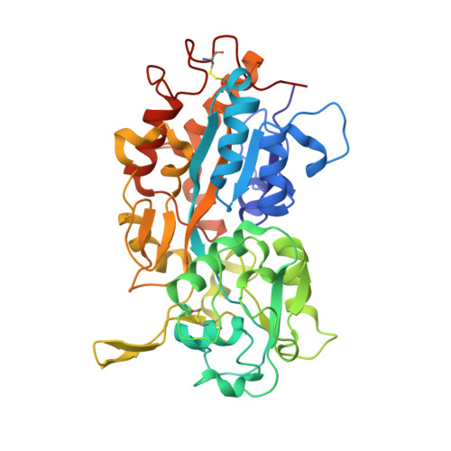
    4M1V

  • PubMed Abstract: 

    Stable and soluble proteins are ideal candidates for functional and structural studies. Unfortunately, some proteins or enzymes can be difficult to isolate, being sometimes poorly expressed in heterologous systems, insoluble and/or unstable. Numerous methods have been developed to address these issues, from the screening of various expression systems to the modification of the target protein itself. Here we use a hydrophobic, aggregation-prone, phosphate-binding protein (HPBP) as a case study. We describe a simple and fast method that selectively uses ancestral mutations to generate a soluble, stable and functional variant of the target protein, here named sHPBP. This variant is highly expressed in Escherichia coli, is easily purified and its structure was solved at much higher resolution than its wild-type progenitor (1.3 versus 1.9 Å, respectively).


  • Organizational Affiliation

    URMITE UMR CNRS-IRD 6236, IFR48, Faculté de Médecine et de Pharmacie, Université de la Méditerranée, Marseille, France.


Macromolecules
Find similar proteins by:  (by identity cutoff)  |  3D Structure
Entity ID: 1
MoleculeChains Sequence LengthOrganismDetailsImage
Phosphate-binding protein378unidentified prokaryotic organismMutation(s): 22 
UniProt
Find proteins for P85173 (Unknown prokaryotic organism)
Explore P85173 
Go to UniProtKB:  P85173
Entity Groups  
Sequence Clusters30% Identity50% Identity70% Identity90% Identity95% Identity100% Identity
UniProt GroupP85173
Sequence Annotations
Expand
  • Reference Sequence
Small Molecules
Ligands 4 Unique
IDChains Name / Formula / InChI Key2D Diagram3D Interactions
PO4
Query on PO4

Download Ideal Coordinates CCD File 
B [auth A]PHOSPHATE ION
O4 P
NBIIXXVUZAFLBC-UHFFFAOYSA-K
GOL
Query on GOL

Download Ideal Coordinates CCD File 
H [auth A]GLYCEROL
C3 H8 O3
PEDCQBHIVMGVHV-UHFFFAOYSA-N
EDO
Query on EDO

Download Ideal Coordinates CCD File 
D [auth A]
E [auth A]
F [auth A]
G [auth A]
I [auth A]
D [auth A],
E [auth A],
F [auth A],
G [auth A],
I [auth A],
J [auth A]
1,2-ETHANEDIOL
C2 H6 O2
LYCAIKOWRPUZTN-UHFFFAOYSA-N
ACT
Query on ACT

Download Ideal Coordinates CCD File 
C [auth A]ACETATE ION
C2 H3 O2
QTBSBXVTEAMEQO-UHFFFAOYSA-M
Experimental Data & Validation

Experimental Data

  • Method: X-RAY DIFFRACTION
  • Resolution: 1.30 Å
  • R-Value Free: 0.143 
  • R-Value Work: 0.107 
  • R-Value Observed: 0.109 
  • Space Group: C 1 2 1
Unit Cell:
Length ( Å )Angle ( ˚ )
a = 125.04α = 90
b = 71.99β = 103.12
c = 38.98γ = 90
Software Package:
Software NamePurpose
MOLREPphasing
REFMACrefinement
XDSdata reduction
XSCALEdata scaling

Structure Validation

View Full Validation Report


Entry History 

Deposition Data

Revision History  (Full details and data files)

  • Version 1.0: 2014-01-01
    Type: Initial release
  • Version 1.1: 2014-03-26
    Changes: Database references
  • Version 1.2: 2023-09-20
    Changes: Data collection, Database references, Derived calculations, Refinement description