4LXF

Crystal structure of M. tuberculosis TreS


Experimental Data Snapshot

  • Method: X-RAY DIFFRACTION
  • Resolution: 2.60 Å
  • R-Value Free: 0.240 
  • R-Value Work: 0.213 
  • R-Value Observed: 0.215 

wwPDB Validation   3D Report Full Report


This is version 1.3 of the entry. See complete history


Literature

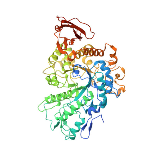
Synthesis of alpha-glucan in mycobacteria involves a hetero-octameric complex of trehalose synthase TreS and Maltokinase Pep2.

Roy, R.Usha, V.Kermani, A.Scott, D.J.Hyde, E.I.Besra, G.S.Alderwick, L.J.Futterer, K.

(2013) ACS Chem Biol 8: 2245-2255

  • DOI: https://doi.org/10.1021/cb400508k
  • Primary Citation of Related Structures:  
    4LXF

  • PubMed Abstract: 

    Recent evidence established that the cell envelope of Mycobacterium tuberculosis, the bacillus causing tuberculosis (TB), is coated by an α-glucan-containing capsule that has been implicated in persistence in a mouse infection model. As one of three known metabolic routes to α-glucan in mycobacteria, the cytoplasmic GlgE-pathway converts trehalose to α(1 → 4),α(1 → 6)-linked glucan in 4 steps. Whether individual reaction steps, catalyzed by trehalose synthase TreS, maltokinase Pep2, and glycosyltransferases GlgE and GlgB, occur independently or in a coordinated fashion is not known. Here, we report the crystal structure of M. tuberculosis TreS, and show by small-angle X-ray scattering and analytical ultracentrifugation that TreS forms tetramers in solution. Together with Pep2, TreS forms a hetero-octameric complex, and we demonstrate that complex formation markedly accelerates maltokinase activity of Pep2. Thus, complex formation may act as part of a regulatory mechanism of the GlgE pathway, which overall must avoid accumulation of toxic pathway intermediates, such as maltose-1-phosphate, and optimize the use of scarce nutrients.


  • Organizational Affiliation

    School of Biosciences, University of Birmingham , Edgbaston, Birmingham B15 2TT, U.K.


Macromolecules
Find similar proteins by:  (by identity cutoff)  |  3D Structure
Entity ID: 1
MoleculeChains Sequence LengthOrganismDetailsImage
Trehalose synthase
A, B
620Mycobacterium tuberculosis H37RvMutation(s): 0 
Gene Names: Rv0126RVBD_0126
EC: 5.4.99.16
Entity Groups  
Sequence Clusters30% Identity50% Identity70% Identity90% Identity95% Identity100% Identity
Sequence Annotations
Expand
  • Reference Sequence
Small Molecules
Ligands 3 Unique
IDChains Name / Formula / InChI Key2D Diagram3D Interactions
SO4
Query on SO4

Download Ideal Coordinates CCD File 
H [auth A]
I [auth A]
J [auth A]
M [auth B]
N [auth B]
H [auth A],
I [auth A],
J [auth A],
M [auth B],
N [auth B],
O [auth B]
SULFATE ION
O4 S
QAOWNCQODCNURD-UHFFFAOYSA-L
GOL
Query on GOL

Download Ideal Coordinates CCD File 
D [auth A],
E [auth A],
F [auth A],
G [auth A],
L [auth B]
GLYCEROL
C3 H8 O3
PEDCQBHIVMGVHV-UHFFFAOYSA-N
CA
Query on CA

Download Ideal Coordinates CCD File 
C [auth A],
K [auth B]
CALCIUM ION
Ca
BHPQYMZQTOCNFJ-UHFFFAOYSA-N
Experimental Data & Validation

Experimental Data

  • Method: X-RAY DIFFRACTION
  • Resolution: 2.60 Å
  • R-Value Free: 0.240 
  • R-Value Work: 0.213 
  • R-Value Observed: 0.215 
  • Space Group: P 32 1 2
Unit Cell:
Length ( Å )Angle ( ˚ )
a = 161.64α = 90
b = 161.64β = 90
c = 139.11γ = 120
Software Package:
Software NamePurpose
ADSCdata collection
PHASERphasing
REFMACrefinement
XDSdata reduction
XSCALEdata scaling

Structure Validation

View Full Validation Report


Entry History 

Deposition Data

Revision History  (Full details and data files)

  • Version 1.0: 2013-08-21
    Type: Initial release
  • Version 1.1: 2013-09-11
    Changes: Database references
  • Version 1.2: 2013-12-25
    Changes: Database references
  • Version 1.3: 2023-09-20
    Changes: Data collection, Database references, Derived calculations, Refinement description