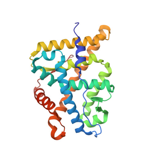
X-ray crystal structure of the ancestral 3-ketosteroid receptor-progesterone-mifepristone complex shows mifepristone bound at the coactivator binding interface.
Colucci, J.K., Ortlund, E.A.(2013) PLoS One 8: e80761-e80761
- PubMed: 24260475
- DOI: https://doi.org/10.1371/journal.pone.0080761
- Primary Citation of Related Structures:
4LTW - PubMed Abstract:
Steroid receptors are a subfamily of nuclear receptors found throughout all metazoans. They are highly important in the regulation of development, inflammation, and reproduction and their misregulation has been implicated in hormone insensitivity syndromes and cancer. Steroid binding to SRs drives a conformational change in the ligand binding domain that promotes nuclear localization and subsequent interaction with coregulator proteins to affect gene regulation. SRs are important pharmaceutical targets, yet most SR-targeting drugs have off-target pharmacology leading to unwanted side effects. A better understanding of the structural mechanisms dictating ligand specificity and the evolution of the forces that created the SR-hormone pairs will enable the design of better pharmaceutical ligands. In order to investigate this relationship, we attempted to crystallize the ancestral 3-ketosteroid receptor (ancSR2) with mifepristone, a SR antagonist. Here, we present the x-ray crystal structure of the ancestral 3-keto steroid receptor (ancSR2)-progesterone complex at a resolution of 2.05 Å. This improves upon our previously reported structure of the ancSR2-progesterone complex, permitting unambiguous assignment of the ligand conformation within the binding pocket. Surprisingly, we find mifepristone, fortuitously docked at the protein surface, poised to interfere with coregulator binding. Recent attention has been given to generating pharmaceuticals that block the coregulator binding site in order to obstruct coregulator binding and achieve tissue-specific SR regulation independent of hormone binding. Mifepristone's interaction with the coactivator cleft of this SR suggests that it may be a useful molecular scaffold for further coactivator binding inhibitor development.
Organizational Affiliation:
Department of Biochemistry and Winship Cancer Institute, Emory University School of Medicine, Atlanta, Georgia, United States of America.