4LS8

Crystal structure of Bacillus subtilis beta-ketoacyl-ACP synthase II (FabF) in a covalent complex with cerulenin


Experimental Data Snapshot

  • Method: X-RAY DIFFRACTION
  • Resolution: 2.10 Å
  • R-Value Free: 0.186 
  • R-Value Work: 0.162 
  • R-Value Observed: 0.162 

wwPDB Validation   3D Report Full Report


Ligand Structure Quality Assessment 


This is version 1.1 of the entry. See complete history


Literature

Structural insights into bacterial resistance to cerulenin.

Trajtenberg, F.Altabe, S.Larrieux, N.Ficarra, F.de Mendoza, D.Buschiazzo, A.Schujman, G.E.

(2014) FEBS J 281: 2324-2338

  • DOI: https://doi.org/10.1111/febs.12785
  • Primary Citation of Related Structures:  
    4LS5, 4LS6, 4LS7, 4LS8

  • PubMed Abstract: 

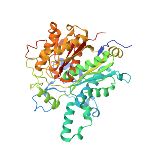
    Cerulenin is a fungal toxin that inhibits both eukaryotic and prokaryotic ketoacyl-acyl carrier protein synthases or condensing enzymes. It has been used experimentally to treat cancer and obesity, and is a potent inhibitor of bacterial growth. Understanding the molecular mechanisms of resistance to cerulenin and similar compounds is thus highly relevant for human health. We have previously described a Bacillus subtilis cerulenin-resistant strain, expressing a point-mutated condensing enzyme FabF (FabF[I108F]) (i.e. FabF with isoleucine 108 substituted by phenylalanine). We now report the crystal structures of wild-type FabF from B. subtilis, both alone and in complex with cerulenin, as well as of the FabF[I108F] mutant protein. The three-dimensional structure of FabF[I108F] constitutes the first atomic model of a condensing enzyme that remains active in the presence of the inhibitor. Soaking the mycotoxin into preformed wild-type FabF crystals allowed for noncovalent binding into its specific pocket within the FabF core. Interestingly, only co-crystallization experiments allowed us to trap the covalent complex. Our structure shows that the covalent bond between Cys163 and cerulenin, in contrast to that previously proposed, implicates carbon C3 of the inhibitor. The similarities between Escherichia coli and B. subtilis FabF structures did not explain the reported inability of ecFabF[I108F] (i.e. FabF from Escherichia coli with isoleucine 108 substituted by phenylalanine) to elongate medium and long-chain acyl-ACPs. We now demonstrate that the E. coli modified enzyme efficiently catalyzes the synthesis of medium and long-chain ketoacyl-ACPs. We also characterized another cerulenin-insensitive form of FabF, conferring a different phenotype in B. subtilis. The structural, biochemical and physiological data presented, shed light on the mechanisms of FabF catalysis and resistance to cerulenin.


  • Organizational Affiliation

    Institut Pasteur de Montevideo, Unit of Protein Crystallography, Montevideo, Uruguay.


Macromolecules
Find similar proteins by:  (by identity cutoff)  |  3D Structure
Entity ID: 1
MoleculeChains Sequence LengthOrganismDetailsImage
3-oxoacyl-[acyl-carrier-protein] synthase 2
A, B
426Bacillus subtilis subsp. subtilis str. 168Mutation(s): 0 
Gene Names: fabFyjaYBSU11340
EC: 2.3.1.179
UniProt
Find proteins for O34340 (Bacillus subtilis (strain 168))
Explore O34340 
Go to UniProtKB:  O34340
Entity Groups  
Sequence Clusters30% Identity50% Identity70% Identity90% Identity95% Identity100% Identity
UniProt GroupO34340
Sequence Annotations
Expand
  • Reference Sequence
Small Molecules
Ligands 4 Unique
IDChains Name / Formula / InChI Key2D Diagram3D Interactions
1XG
Query on 1XG

Download Ideal Coordinates CCD File 
C [auth A],
J [auth B]
(3R,7E,10E)-3-hydroxy-4-oxododeca-7,10-dienamide
C12 H19 N O3
QEPYZBPOTYDXNA-IZFUEKCOSA-N
EDO
Query on EDO

Download Ideal Coordinates CCD File 
F [auth A],
G [auth A],
I [auth A]
1,2-ETHANEDIOL
C2 H6 O2
LYCAIKOWRPUZTN-UHFFFAOYSA-N
CL
Query on CL

Download Ideal Coordinates CCD File 
E [auth A]CHLORIDE ION
Cl
VEXZGXHMUGYJMC-UHFFFAOYSA-M
NA
Query on NA

Download Ideal Coordinates CCD File 
D [auth A],
H [auth A],
K [auth B]
SODIUM ION
Na
FKNQFGJONOIPTF-UHFFFAOYSA-N
Experimental Data & Validation

Experimental Data

  • Method: X-RAY DIFFRACTION
  • Resolution: 2.10 Å
  • R-Value Free: 0.186 
  • R-Value Work: 0.162 
  • R-Value Observed: 0.162 
  • Space Group: P 21 21 21
Unit Cell:
Length ( Å )Angle ( ˚ )
a = 86.44α = 90
b = 87.56β = 90
c = 116.44γ = 90
Software Package:
Software NamePurpose
MOSFLMdata reduction
SCALAdata scaling
BUSTER-TNTrefinement
PDB_EXTRACTdata extraction
MAR345dtbdata collection
BUSTERrefinement

Structure Validation

View Full Validation Report


Ligand Structure Quality Assessment 


Entry History 

Deposition Data

Revision History  (Full details and data files)

  • Version 1.0: 2014-04-02
    Type: Initial release
  • Version 1.1: 2014-06-04
    Changes: Database references