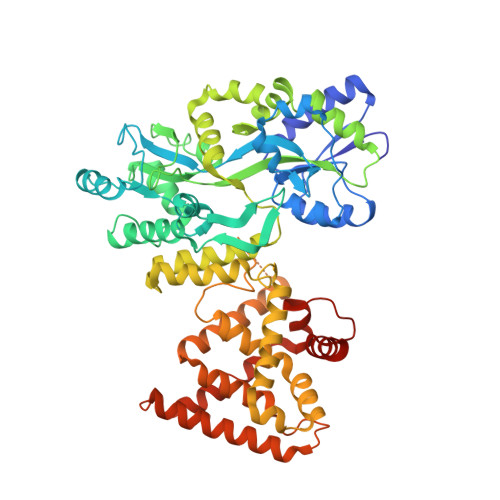
The Crystal Structure of the Orphan Nuclear Receptor NR2E3/PNR Ligand Binding Domain Reveals a Dimeric Auto-Repressed Conformation.
Tan, M.H., Zhou, X.E., Soon, F.F., Li, X., Li, J., Yong, E.L., Melcher, K., Xu, H.E.(2013) PLoS One 8: e74359-e74359
- PubMed: 24069298
- DOI: https://doi.org/10.1371/journal.pone.0074359
- Primary Citation of Related Structures:
4LOG - PubMed Abstract:
Photoreceptor-specific nuclear receptor (PNR, NR2E3) is a key transcriptional regulator of human photoreceptor differentiation and maintenance. Mutations in the NR2E3-encoding gene cause various retinal degenerations, including Enhanced S-cone syndrome, retinitis pigmentosa, and Goldman-Favre disease. Although physiological ligands have not been identified, it is believed that binding of small molecule agonists, receptor desumoylation, and receptor heterodimerization may switch NR2E3 from a transcriptional repressor to an activator. While these features make NR2E3 a potential therapeutic target for the treatment of retinal diseases, there has been a clear lack of structural information for the receptor. Here, we report the crystal structure of the apo NR2E3 ligand binding domain (LBD) at 2.8 Å resolution. Apo NR2E3 functions as transcriptional repressor in cells and the structure of its LBD is in a dimeric auto-repressed conformation. In this conformation, the putative ligand binding pocket is filled with bulky hydrophobic residues and the activation-function-2 (AF2) helix occupies the canonical cofactor binding site. Mutations designed to disrupt either the AF2/cofactor-binding site interface or the dimer interface compromised the transcriptional repressor activity of this receptor. Together, these results reveal several conserved structural features shared by related orphan nuclear receptors, suggest that most disease-causing mutations affect the receptor's structural integrity, and allowed us to model a putative active conformation that can accommodate small ligands in its pocket.
Organizational Affiliation:
Laboratory of Structural Sciences, Van Andel Research Institute, Grand Rapids, Michigan, United States of America ; Department of Obstetrics & Gynecology, National University Hospital, Yong Loo Lin School of Medicine, National University of Singapore, Singapore, Singapore.