4LKU

Structure of the C-terminal domain of the E. coli mechanosensitive channel of large conductance


Experimental Data Snapshot

  • Method: X-RAY DIFFRACTION
  • Resolution: 1.45 Å
  • R-Value Free: 0.225 
  • R-Value Work: 0.169 
  • R-Value Observed: 0.172 

wwPDB Validation   3D Report Full Report


This is version 1.3 of the entry. See complete history


Literature

Structure and stability of the C-terminal helical bundle of the E. coli mechanosensitive channel of large conductance.

Walton, T.A.Rees, D.C.

(2013) Protein Sci 22: 1592-1601

  • DOI: https://doi.org/10.1002/pro.2360
  • Primary Citation of Related Structures:  
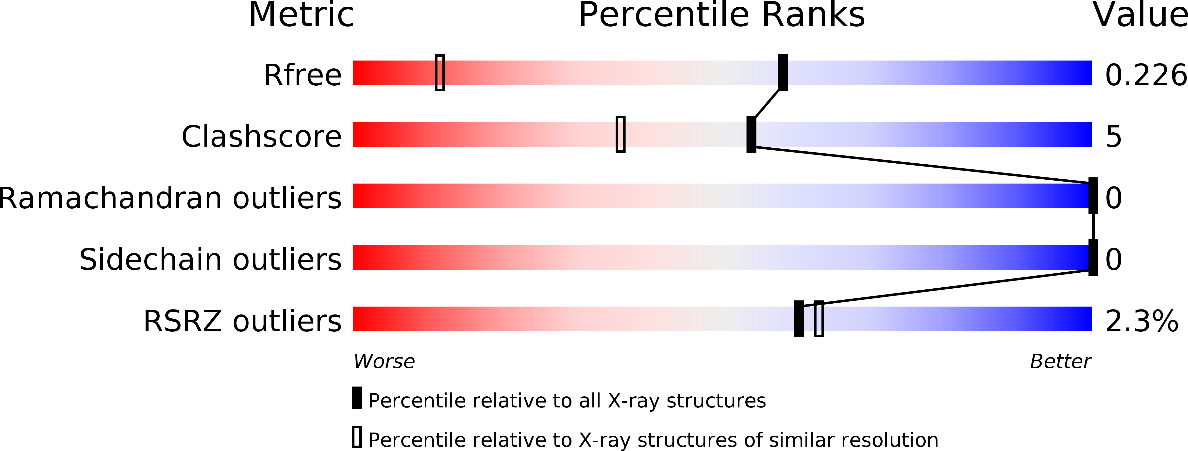
    4LKU

  • PubMed Abstract: 

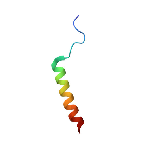
    The crystal structure of the cytoplasmic domain (CTD) from the mechanosensitive channel of large conductance (MscL) in E. coli has been determined at 1.45 Å resolution. This domain forms a pentameric coiled coil similar to that observed in the structure of MscL from M. tuberculosis and also found in the cartilage oligomeric matrix protein (COMPcc). It contains canonical hydrophobic and atypical ionic interactions compared to previously characterized coiled coil structures. Thermodynamic analysis indicates that while the free EcMscL-CTD is less stable than other coiled coils, it is likely to remain folded in context of the full-length channel.


  • Organizational Affiliation

    Division of Chemistry and Chemical Engineering, Howard Hughes Medical Institute, California Institute of Technology, Pasadena, California, 91125.


Macromolecules
Find similar proteins by:  (by identity cutoff)  |  3D Structure
Entity ID: 1
MoleculeChains Sequence LengthOrganismDetailsImage
Large-conductance mechanosensitive channel
A, B, C, D, E
29Escherichia coli DH1Mutation(s): 0 
UniProt
Find proteins for P0A742 (Escherichia coli (strain K12))
Explore P0A742 
Go to UniProtKB:  P0A742
Entity Groups  
Sequence Clusters30% Identity50% Identity70% Identity90% Identity95% Identity100% Identity
UniProt GroupP0A742
Sequence Annotations
Expand
  • Reference Sequence
Experimental Data & Validation

Experimental Data

  • Method: X-RAY DIFFRACTION
  • Resolution: 1.45 Å
  • R-Value Free: 0.225 
  • R-Value Work: 0.169 
  • R-Value Observed: 0.172 
  • Space Group: P 1 21 1
Unit Cell:
Length ( Å )Angle ( ˚ )
a = 32.25α = 90
b = 55.12β = 93.58
c = 32.85γ = 90
Software Package:
Software NamePurpose
SCALAdata scaling
PHASERphasing
PHENIXrefinement
PDB_EXTRACTdata extraction
Blu-Icedata collection
MOSFLMdata reduction

Structure Validation

View Full Validation Report


Entry History 

Deposition Data

Revision History  (Full details and data files)

  • Version 1.0: 2013-10-02
    Type: Initial release
  • Version 1.1: 2013-11-20
    Changes: Database references
  • Version 1.2: 2017-11-15
    Changes: Refinement description
  • Version 1.3: 2024-02-28
    Changes: Data collection, Database references