


Crystal structure of a enoyl-CoA hydratase from Mycobacterium avium subsp. paratuberculosis K-10
Kumaran, D., Almo, S.C., Swaminathan, S.To be published.
Experimental Data Snapshot
wwPDB Validation 3D Report Full Report

Entity ID: 1 | |||||
|---|---|---|---|---|---|
| Molecule | Chains | Sequence Length | Organism | Details | Image |
| enoyl-CoA hydratase | 292 | Mycobacterium avium subsp. paratuberculosis K-10 | Mutation(s): 0 Gene Names: echA11, MAP_2639 | 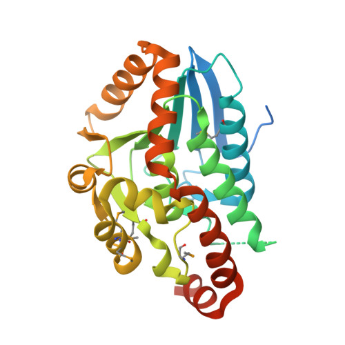 | |
UniProt | |||||
Find proteins for Q73WM1 (Mycolicibacterium paratuberculosis (strain ATCC BAA-968 / K-10)) Explore Q73WM1 Go to UniProtKB: Q73WM1 | |||||
Entity Groups | |||||
| Sequence Clusters | 30% Identity50% Identity70% Identity90% Identity95% Identity100% Identity | ||||
| UniProt Group | Q73WM1 | ||||
Sequence AnnotationsExpand | |||||
| |||||
| Modified Residues 1 Unique | |||||
|---|---|---|---|---|---|
| ID | Chains | Type | Formula | 2D Diagram | Parent |
| MSE Query on MSE | A, B, C | L-PEPTIDE LINKING | C5 H11 N O2 Se |  | MET |
| Length ( Å ) | Angle ( ˚ ) |
|---|---|
| a = 78.328 | α = 90 |
| b = 78.328 | β = 90 |
| c = 212.933 | γ = 120 |
| Software Name | Purpose |
|---|---|
| CBASS | data collection |
| SHELXD | phasing |
| SHELXE | model building |
| ARP/wARP | model building |
| Coot | model building |
| REFMAC | refinement |
| HKL-2000 | data reduction |
| HKL-2000 | data scaling |
RCSB PDB (citation) is hosted by
RCSB PDB is a member of the

