The C2B Domain Is the Primary Ca(2+) Sensor in DOC2B: A Structural and Functional Analysis.
Giladi, M., Michaeli, L., Almagor, L., Bar-On, D., Buki, T., Ashery, U., Khananshvili, D., Hirsch, J.A.(2013) J Mol Biol 425: 4629-4641
- PubMed: 23994332
- DOI: https://doi.org/10.1016/j.jmb.2013.08.017
- Primary Citation of Related Structures:
4LCV, 4LDC - PubMed Abstract:
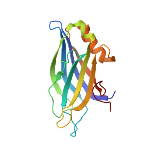
DOC2B (double-C2 domain) protein is thought to be a high-affinity Ca(2+) sensor for spontaneous and asynchronous neurotransmitter release. To elucidate the molecular features underlying its physiological role, we determined the crystal structures of its isolated C2A and C2B domains and examined their Ca(2+)-binding properties. We further characterized the solution structure of the tandem domains (C2AB) using small-angle X-ray scattering. In parallel, we tested structure-function correlates with live cell imaging tools. We found that, despite striking structural similarity, C2B binds Ca(2+) with considerably higher affinity than C2A. The C2AB solution structure is best modeled as two domains with a highly flexible orientation and no difference in the presence or absence of Ca(2+). In addition, kinetic studies of C2AB demonstrate that, in the presence of unilamellar vesicles, Ca(2+) binding is stabilized, as reflected by the ~10-fold slower rate of Ca(2+) dissociation than in the absence of vesicles. In cells, isolated C2B translocates to the plasma membrane (PM) with an EC50 of 400 nM while the C2A does not translocate at submicromolar Ca(2+) concentrations, supporting the biochemical observations. Nevertheless, C2AB translocates to the PM with an ~2-fold lower EC50 and to a greater extent than C2B. Our results, together with previous studies, reveal that the C2B is the primary Ca(2+) sensing unit in DOC2B, whereas C2A enhances the interaction of C2AB with the PM.
Organizational Affiliation:
Department of Physiology and Pharmacology, Sackler School of Medicine, Tel-Aviv University, Ramat-Aviv 69978, Israel.