


Crystal structure of PWWP domain of human PWWP Domain-Containing Protein 2B
Dombrovski, L., Dong, A., Tempel, W., Loppnau, P., Bountra, C., Arrowsmith, C.H., Edwards, A.M., Brown, P.J., Wu, H., Structural Genomics Consortium (SGC)To be published.
Experimental Data Snapshot
wwPDB Validation 3D Report Full Report

Entity ID: 1 | |||||
|---|---|---|---|---|---|
| Molecule | Chains | Sequence Length | Organism | Details | Image |
| PWWP domain-containing protein 2B | 117 | Homo sapiens | Mutation(s): 0 Gene Names: PWWP2B, PWWP2 | 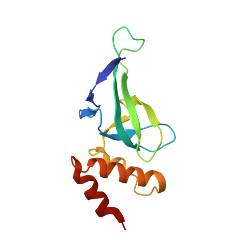 | |
UniProt & NIH Common Fund Data Resources | |||||
Find proteins for Q6NUJ5 (Homo sapiens) Explore Q6NUJ5 Go to UniProtKB: Q6NUJ5 | |||||
PHAROS: Q6NUJ5 GTEx: ENSG00000171813 | |||||
Entity Groups | |||||
| Sequence Clusters | 30% Identity50% Identity70% Identity90% Identity95% Identity100% Identity | ||||
| UniProt Group | Q6NUJ5 | ||||
Sequence AnnotationsExpand | |||||
| |||||
| Ligands 2 Unique | |||||
|---|---|---|---|---|---|
| ID | Chains | Name / Formula / InChI Key | 2D Diagram | 3D Interactions | |
| GOL Query on GOL | B [auth A] | GLYCEROL C3 H8 O3 PEDCQBHIVMGVHV-UHFFFAOYSA-N |  | ||
| UNX Query on UNX | C [auth A] D [auth A] E [auth A] F [auth A] G [auth A] | UNKNOWN ATOM OR ION X |  | ||
| Length ( Å ) | Angle ( ˚ ) |
|---|---|
| a = 32.148 | α = 90 |
| b = 41.055 | β = 90 |
| c = 77.972 | γ = 90 |
| Software Name | Purpose |
|---|---|
| REFMAC | refinement |
| PDB_EXTRACT | data extraction |
| JBluIce-EPICS | data collection |
| HKL-3000 | data reduction |
| HKL-3000 | data scaling |
| SOLVE | phasing |
RCSB PDB (citation) is hosted by
RCSB PDB is a member of the

