4LCE

CtBP1 in complex with substrate MTOB


Experimental Data Snapshot

  • Method: X-RAY DIFFRACTION
  • Resolution: 2.38 Å
  • R-Value Free: 0.239 
  • R-Value Work: 0.200 
  • R-Value Observed: 0.202 

wwPDB Validation   3D Report Full Report


Ligand Structure Quality Assessment 


This is version 1.5 of the entry. See complete history


Literature

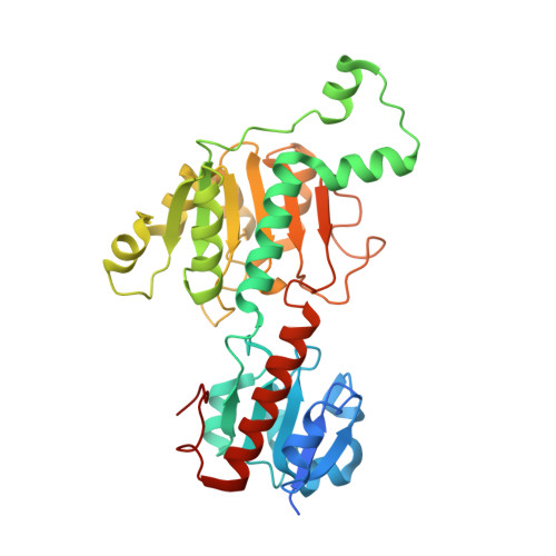
Crystal structures of human CtBP in complex with substrate MTOB reveal active site features useful for inhibitor design.

Hilbert, B.J.Grossmann, S.R.Schiffer, C.A.Royer, W.E.

(2014) FEBS Lett 588: 1743-1748

  • DOI: https://doi.org/10.1016/j.febslet.2014.03.026
  • Primary Citation of Related Structures:  
    4LCE, 4LCJ

  • PubMed Abstract: 

    The oncogenic corepressors C-terminal Binding Protein (CtBP) 1 and 2 harbor regulatory d-isomer specific 2-hydroxyacid dehydrogenase (d2-HDH) domains. 4-Methylthio 2-oxobutyric acid (MTOB) exhibits substrate inhibition and can interfere with CtBP oncogenic activity in cell culture and mice. Crystal structures of human CtBP1 and CtBP2 in complex with MTOB and NAD(+) revealed two key features: a conserved tryptophan that likely contributes to substrate specificity and a hydrophilic cavity that links MTOB with an NAD(+) phosphate. Neither feature is present in other d2-HDH enzymes. These structures thus offer key opportunities for the development of highly selective anti-neoplastic CtBP inhibitors.


  • Organizational Affiliation

    Department of Biochemistry and Molecular Pharmacology, University of Massachusetts Medical School, Worcester, MA 01605, USA.


Macromolecules
Find similar proteins by:  (by identity cutoff)  |  3D Structure
Entity ID: 1
MoleculeChains Sequence LengthOrganismDetailsImage
C-terminal-binding protein 1347Homo sapiensMutation(s): 0 
Gene Names: CTBPCTBP1
EC: 1.1.1
UniProt & NIH Common Fund Data Resources
Find proteins for Q13363 (Homo sapiens)
Explore Q13363 
Go to UniProtKB:  Q13363
PHAROS:  Q13363
GTEx:  ENSG00000159692 
Entity Groups  
Sequence Clusters30% Identity50% Identity70% Identity90% Identity95% Identity100% Identity
UniProt GroupQ13363
Sequence Annotations
Expand
  • Reference Sequence
Small Molecules
Ligands 2 Unique
IDChains Name / Formula / InChI Key2D Diagram3D Interactions
NAD
Query on NAD

Download Ideal Coordinates CCD File 
B [auth A]NICOTINAMIDE-ADENINE-DINUCLEOTIDE
C21 H27 N7 O14 P2
BAWFJGJZGIEFAR-NNYOXOHSSA-N
KMT
Query on KMT

Download Ideal Coordinates CCD File 
C [auth A]4-(METHYLSULFANYL)-2-OXOBUTANOIC ACID
C5 H8 O3 S
SXFSQZDSUWACKX-UHFFFAOYSA-N
Binding Affinity Annotations 
IDSourceBinding Affinity
KMT BindingDB:  4LCE Kd: min: 1.26e+6, max: 2.96e+6 (nM) from 2 assay(s)
IC50: 3.00e+5 (nM) from 1 assay(s)
-TΔS: 32.25 (kJ/mol) from 1 assay(s)
ΔH: min: -4.73e+1, max: -1.40e+1 (kJ/mol) from 2 assay(s)
ΔG: min: -1.60e+1, max: -1.40e+1 (kJ/mol) from 2 assay(s)
Experimental Data & Validation

Experimental Data

  • Method: X-RAY DIFFRACTION
  • Resolution: 2.38 Å
  • R-Value Free: 0.239 
  • R-Value Work: 0.200 
  • R-Value Observed: 0.202 
  • Space Group: P 64 2 2
Unit Cell:
Length ( Å )Angle ( ˚ )
a = 88.97α = 90
b = 88.97β = 90
c = 161.538γ = 120
Software Package:
Software NamePurpose
DENZOdata reduction
SCALEPACKdata scaling
PHASERphasing
REFMACrefinement
PDB_EXTRACTdata extraction
ADSCdata collection
HKL-2000data reduction
HKL-2000data scaling

Structure Validation

View Full Validation Report


Ligand Structure Quality Assessment 


Entry History 

Deposition Data

Revision History  (Full details and data files)

  • Version 1.0: 2014-03-19
    Type: Initial release
  • Version 1.1: 2014-04-09
    Changes: Database references
  • Version 1.2: 2014-05-14
    Changes: Database references
  • Version 1.3: 2014-11-12
    Changes: Structure summary
  • Version 1.4: 2017-11-15
    Changes: Refinement description
  • Version 1.5: 2024-02-28
    Changes: Data collection, Database references, Derived calculations