4LAG

Structure-Based Design of New Dihydrofolate Reductase Antibacterial Agents: 7-(Benzimidazol-1-yl)-2,4-diaminoquinazolines


Experimental Data Snapshot

  • Method: X-RAY DIFFRACTION
  • Resolution: 1.70 Å
  • R-Value Free: 0.214 
  • R-Value Work: 0.196 
  • R-Value Observed: 0.197 

wwPDB Validation   3D Report Full Report


Ligand Structure Quality Assessment 


This is version 1.2 of the entry. See complete history


Literature

Structure-Based Design of New Dihydrofolate Reductase Antibacterial Agents: 7-(Benzimidazol-1-yl)-2,4-diaminoquinazolines.

Lam, T.Hilgers, M.T.Cunningham, M.L.Kwan, B.P.Nelson, K.J.Brown-Driver, V.Ong, V.Trzoss, M.Hough, G.Shaw, K.J.Finn, J.

(2014) J Med Chem 57: 651-668

  • DOI: https://doi.org/10.1021/jm401204g
  • Primary Citation of Related Structures:  
    4LAE, 4LAG, 4LAH, 4LEK

  • PubMed Abstract: 

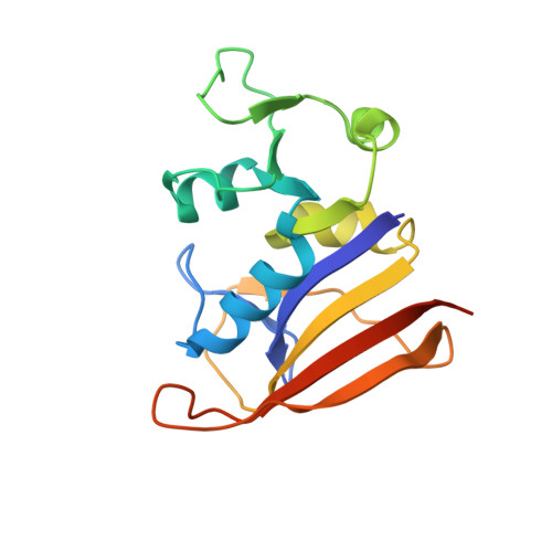
    A new series of dihydrofolate reductase (DHFR) inhibitors, the 7-(benzimidazol-1-yl)-2,4-diaminoquinazolines, were designed and optimized for antibacterial potency and enzyme selectivity. The most potent inhibitors in this series contained a five-membered heterocycle at the 2-position of the benzimidazole, leading to highly potent and selective compounds that exploit the differences in the size of a binding pocket adjacent to the NADPH cofactor between the bacterial and human DHFR enzymes. Typical of these compounds is 7-((2-thiazol-2-yl)benzimidazol-1-yl)-2,4 diaminoquinazoline, which is a potent inhibitor of S. aureus DHFR (Ki = 0.002 nM) with 46700-fold selectivity over human DHFR. This compound also has high antibacterial potency on Gram-positive bacteria with an MIC versus wild type S. aureus of 0.0125 μg/mL and a MIC versus trimethoprim-resistant S. aureus of 0.25 μg/mL. In vivo efficacy versus a S. aureus septicemia was demonstrated, highlighting the potential of this new series.


  • Organizational Affiliation

    Trius Therapeutics Inc. , 6310 Nancy Ridge Drive, San Diego, California 92121, United States.


Macromolecules
Find similar proteins by:  (by identity cutoff)  |  3D Structure
Entity ID: 1
MoleculeChains Sequence LengthOrganismDetailsImage
Dihydrofolate reductaseA [auth X]167Staphylococcus aureusMutation(s): 0 
Gene Names: folA
EC: 1.5.1.3
UniProt
Find proteins for P0A017 (Staphylococcus aureus)
Explore P0A017 
Go to UniProtKB:  P0A017
Entity Groups  
Sequence Clusters30% Identity50% Identity70% Identity90% Identity95% Identity100% Identity
UniProt GroupP0A017
Sequence Annotations
Expand
  • Reference Sequence
Small Molecules
Ligands 2 Unique
IDChains Name / Formula / InChI Key2D Diagram3D Interactions
NAP
Query on NAP

Download Ideal Coordinates CCD File 
B [auth X]NADP NICOTINAMIDE-ADENINE-DINUCLEOTIDE PHOSPHATE
C21 H28 N7 O17 P3
XJLXINKUBYWONI-NNYOXOHSSA-N
1VN
Query on 1VN

Download Ideal Coordinates CCD File 
C [auth X]6-chloro-7-[5,6-dimethyl-2-(1,3-thiazol-2-yl)-1H-benzimidazol-1-yl]quinazoline-2,4-diamine
C20 H16 Cl N7 S
WSVACSSBUCFLCD-UHFFFAOYSA-N
Binding Affinity Annotations 
IDSourceBinding Affinity
1VN BindingDB:  4LAG Ki: min: 0.01, max: 0.02 (nM) from 2 assay(s)
Binding MOAD:  4LAG Ki: 0.01 (nM) from 1 assay(s)
Experimental Data & Validation

Experimental Data

  • Method: X-RAY DIFFRACTION
  • Resolution: 1.70 Å
  • R-Value Free: 0.214 
  • R-Value Work: 0.196 
  • R-Value Observed: 0.197 
  • Space Group: P 61 2 2
Unit Cell:
Length ( Å )Angle ( ˚ )
a = 79.302α = 90
b = 79.302β = 90
c = 107.206γ = 120
Software Package:
Software NamePurpose
MOSFLMdata reduction
SCALEPACKdata scaling
PHASERphasing
REFMACrefinement
PDB_EXTRACTdata extraction

Structure Validation

View Full Validation Report


Ligand Structure Quality Assessment 


Entry History 

Deposition Data

Revision History  (Full details and data files)

  • Version 1.0: 2014-02-19
    Type: Initial release
  • Version 1.1: 2014-02-26
    Changes: Database references
  • Version 1.2: 2024-02-28
    Changes: Data collection, Database references, Derived calculations