Crystal structure and activity of protein L-isoaspartyl-O-methyltransferase from Vibrio cholerae, and the effect of AdoHcy binding.
Chatterjee, T., Mukherjee, D., Banerjee, M., Chatterjee, B.K., Chakrabarti, P.(2015) Arch Biochem Biophys 583: 140-149
- PubMed: 26255776
- DOI: https://doi.org/10.1016/j.abb.2015.08.001
- Primary Citation of Related Structures:
4L7V - PubMed Abstract:
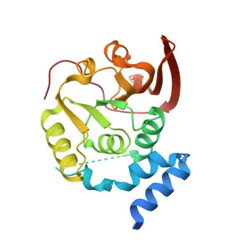
The repair enzyme Protein L-isoaspartyl-O-methyltransferase (PIMT) is widely distributed in various organisms. PIMT catalyzes S-adenosylmethionine (AdoMet) dependent methylation of abnormal L-isoaspartyl residues, formed by the deamidation of asparagines and isomerization of aspartates. We report the crystal structure of PIMT of Vibrio cholerae (VcPIMT), the aetiological agent for cholera, complexed with the demethylated cofactor S-adenosyl-L-homocysteine (AdoHcy) to 2.05 Å resolution. A stretch of residues (39-58), lining the substrate-binding site, is disordered. Urea-induced unfolding free energy for apo and VcPIMT-AdoHcy complex reveals greater stability for the cofactor-bound protein. The kinetic parameters for the methyltransferase activity of the recombinant VcPIMT was determined using a continuous spectrophotometric color-based assay using the peptide substrate [VYP(L-isoD)HA]. The enzyme exhibited activity higher than the Escherichia coli enzyme and closer to those from thermophilic bacteria and the mammalian source. The association constant for substrate binding is 2.29 × 10(6) M(-1), quite similar to that for AdoHcy. The crystal structure and the model of the peptide-bound structure indicate that the majority of the interactions used for cofactor/substrate binding are provided by the main-chain atoms. Evolutionary relationships derived based on a phylogenetic tree constructed using the PIMT sequences are in conformity with the crystal structures of nine AdoHcy-bound PIMTs.
Organizational Affiliation:
Department of Biochemistry, Bose Institute, P1/12, CIT Scheme VIIM, Kolkata 700054, India.