4L67

Crystal Structure of Catalytic Domain of PAK4


Experimental Data Snapshot

  • Method: X-RAY DIFFRACTION
  • Resolution: 2.80 Å
  • R-Value Free: 0.333 
  • R-Value Work: 0.228 
  • R-Value Observed: 0.233 

wwPDB Validation   3D Report Full Report


This is version 1.2 of the entry. See complete history


Literature

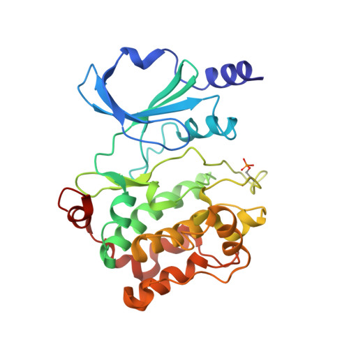
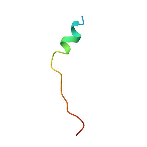
NMR binding and crystal structure reveal that intrinsically-unstructured regulatory domain auto-inhibits PAK4 by a mechanism different for that of PAK1

Wang, W.Lim, L.Baskaran, Y.Manser, E.Song, J.

(2013) Biochem Biophys Res Commun 438: 169-174

  • DOI: https://doi.org/10.1016/j.bbrc.2013.07.047
  • Primary Citation of Related Structures:  
    4L67

  • PubMed Abstract: 

    Six human PAK members are classified into groups I (PAKs 1-3) and II (PAK4-6). Previously, only group I PAKs were thought to be auto-inhibited but very recently PAK4, the prototype of group II PAKs, has also been shown to be auto-inhibited by its N-terminal regulatory domain. However, the complete auto-inhibitory domain (AID) sequence remains undefined and the mechanism underlying its auto-inhibition is largely elusive. Here, the N-terminal regulatory domain of PAK4 sufficient for auto-inhibiting and binding Cdc42/Rac was characterized to be intrinsically unstructured, but nevertheless we identified the entire AID sequence by NMR. Strikingly, an AID peptide was derived by deleting the binding-unnecessary residues, which has a Kd of 320 nM to the PAK4 catalytic domain. Consequently, the PAK4 crystal structure complexed with the entire AID has been determined, which reveals that the complete kinase cleft is occupied by 20 AID residuescomposed of an N-terminal α-helix and a previously-identified pseudosubstrate motif, thus achieving auto-inhibition. Our study reveals that PAK4 is auto-inhibited by a novel mechanism which is completely different from that for PAK1, thus bearing critical implications for design of inhibitors specific for group II PAKs.


  • Organizational Affiliation

    Department of Biological Sciences, Faculty of Science, National University of Singapore, 10 Kent Ridge Crescent, Singapore 119260, Singapore.


Macromolecules
Find similar proteins by:  (by identity cutoff)  |  3D Structure
Entity ID: 1
MoleculeChains Sequence LengthOrganismDetailsImage
Serine/threonine-protein kinase PAK 4292Homo sapiensMutation(s): 0 
Gene Names: PAK4KIAA1142
EC: 2.7.11.1
UniProt & NIH Common Fund Data Resources
Find proteins for O96013 (Homo sapiens)
Explore O96013 
Go to UniProtKB:  O96013
PHAROS:  O96013
GTEx:  ENSG00000130669 
Entity Groups  
Sequence Clusters30% Identity50% Identity70% Identity90% Identity95% Identity100% Identity
UniProt GroupO96013
Sequence Annotations
Expand
  • Reference Sequence

Find similar proteins by:  Sequence   |   3D Structure  

Entity ID: 2
MoleculeChains Sequence LengthOrganismDetailsImage
Serine/threonine-protein kinase PAK 425Homo sapiensMutation(s): 0 
Gene Names: PAK4KIAA1142
EC: 2.7.11.1
UniProt & NIH Common Fund Data Resources
Find proteins for O96013 (Homo sapiens)
Explore O96013 
Go to UniProtKB:  O96013
PHAROS:  O96013
GTEx:  ENSG00000130669 
Entity Groups  
Sequence Clusters30% Identity50% Identity70% Identity90% Identity95% Identity100% Identity
UniProt GroupO96013
Sequence Annotations
Expand
  • Reference Sequence
Small Molecules
Modified Residues  1 Unique
IDChains TypeFormula2D DiagramParent
SEP
Query on SEP
A
L-PEPTIDE LINKINGC3 H8 N O6 PSER
Experimental Data & Validation

Experimental Data

  • Method: X-RAY DIFFRACTION
  • Resolution: 2.80 Å
  • R-Value Free: 0.333 
  • R-Value Work: 0.228 
  • R-Value Observed: 0.233 
  • Space Group: P 41 21 2
Unit Cell:
Length ( Å )Angle ( ˚ )
a = 65.175α = 90
b = 65.175β = 90
c = 184.567γ = 90
Software Package:
Software NamePurpose
CrystalCleardata collection
CCP4model building
REFMACrefinement
HKL-2000data reduction
HKL-2000data scaling
CCP4phasing

Structure Validation

View Full Validation Report


Entry History 

Deposition Data

Revision History  (Full details and data files)

  • Version 1.0: 2013-08-14
    Type: Initial release
  • Version 1.1: 2013-09-04
    Changes: Database references
  • Version 1.2: 2023-11-08
    Changes: Data collection, Database references, Derived calculations, Refinement description