4L57

High resolutin structure of human cytosolic 5'(3')-deoxyribonucleotidase


Experimental Data Snapshot

  • Method: X-RAY DIFFRACTION
  • Resolution: 1.08 Å
  • R-Value Free: 0.183 
  • R-Value Work: 0.144 
  • R-Value Observed: 0.144 

wwPDB Validation   3D Report Full Report


This is version 1.3 of the entry. See complete history


Literature

Structures of human cytosolic and mitochondrial nucleotidases: implications for structure-based design of selective inhibitors.

Pachl, P.Fabry, M.Rosenberg, I.Simak, O.Rezacova, P.Brynda, J.

(2014) Acta Crystallogr D Biol Crystallogr 70: 461-470

  • DOI: https://doi.org/10.1107/S1399004713030502
  • Primary Citation of Related Structures:  
    4L57, 4L6A

  • PubMed Abstract: 

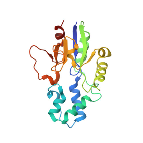
    The human 5'(3')-deoxyribonucleotidases catalyze the dephosphorylation of deoxyribonucleoside monophosphates to the corresponding deoxyribonucleosides and thus help to maintain the balance between pools of nucleosides and nucleotides. Here, the structures of human cytosolic deoxyribonucleotidase (cdN) at atomic resolution (1.08 Å) and mitochondrial deoxyribonucleotidase (mdN) at near-atomic resolution (1.4 Å) are reported. The attainment of an atomic resolution structure allowed interatomic distances to be used to assess the probable protonation state of the phosphate anion and the side chains in the enzyme active site. A detailed comparison of the cdN and mdN active sites allowed the design of a cdN-specific inhibitor.


  • Organizational Affiliation

    Institute of Molecular Genetics, Academy of Sciences of the Czech Republic, v.v.i., Flemingovo nám. 2, 166 37 Prague 6, Czech Republic.


Macromolecules
Find similar proteins by:  (by identity cutoff)  |  3D Structure
Entity ID: 1
MoleculeChains Sequence LengthOrganismDetailsImage
5'(3')-deoxyribonucleotidase, cytosolic type
A, B
195Homo sapiensMutation(s): 0 
Gene Names: DNT1NT5CUMPH2
EC: 3.1.3
UniProt & NIH Common Fund Data Resources
Find proteins for Q8TCD5 (Homo sapiens)
Explore Q8TCD5 
Go to UniProtKB:  Q8TCD5
PHAROS:  Q8TCD5
GTEx:  ENSG00000125458 
Entity Groups  
Sequence Clusters30% Identity50% Identity70% Identity90% Identity95% Identity100% Identity
UniProt GroupQ8TCD5
Sequence Annotations
Expand
  • Reference Sequence
Small Molecules
Ligands 4 Unique
IDChains Name / Formula / InChI Key2D Diagram3D Interactions
PO4
Query on PO4

Download Ideal Coordinates CCD File 
D [auth A],
L [auth B]
PHOSPHATE ION
O4 P
NBIIXXVUZAFLBC-UHFFFAOYSA-K
GOL
Query on GOL

Download Ideal Coordinates CCD File 
E [auth A]
F [auth A]
G [auth A]
H [auth A]
I [auth A]
E [auth A],
F [auth A],
G [auth A],
H [auth A],
I [auth A],
M [auth B]
GLYCEROL
C3 H8 O3
PEDCQBHIVMGVHV-UHFFFAOYSA-N
MG
Query on MG

Download Ideal Coordinates CCD File 
C [auth A],
K [auth B]
MAGNESIUM ION
Mg
JLVVSXFLKOJNIY-UHFFFAOYSA-N
NA
Query on NA

Download Ideal Coordinates CCD File 
J [auth A]SODIUM ION
Na
FKNQFGJONOIPTF-UHFFFAOYSA-N
Experimental Data & Validation

Experimental Data

  • Method: X-RAY DIFFRACTION
  • Resolution: 1.08 Å
  • R-Value Free: 0.183 
  • R-Value Work: 0.144 
  • R-Value Observed: 0.144 
  • Space Group: P 1
Unit Cell:
Length ( Å )Angle ( ˚ )
a = 39.128α = 68.16
b = 46.493β = 81.47
c = 61.409γ = 75.28
Software Package:
Software NamePurpose
SHELXrefinement
PDB_EXTRACTdata extraction
SHELXLrefinement

Structure Validation

View Full Validation Report


Entry History 

Deposition Data

Revision History  (Full details and data files)

  • Version 1.0: 2014-02-12
    Type: Initial release
  • Version 1.1: 2014-03-19
    Changes: Refinement description
  • Version 1.2: 2018-04-18
    Changes: Data collection, Database references
  • Version 1.3: 2024-02-28
    Changes: Data collection, Database references, Derived calculations