4L37

SP2-SP3 - a complex of two storage proteins from Bombyx mori hemolymph


Experimental Data Snapshot

  • Method: X-RAY DIFFRACTION
  • Resolution: 2.90 Å
  • R-Value Free: 0.223 
  • R-Value Work: 0.166 
  • R-Value Observed: 0.167 

wwPDB Validation   3D Report Full Report


This is version 2.1 of the entry. See complete history


Literature

Crystallographic identification of an unexpected protein complex in silkworm haemolymph.

Pietrzyk, A.J.Bujacz, A.Mueller-Dieckmann, J.ochynska, M.Jaskolski, M.Bujacz, G.

(2013) Acta Crystallogr D Biol Crystallogr 69: 2353-2364

  • DOI: https://doi.org/10.1107/S0907444913021823
  • Primary Citation of Related Structures:  
    4L37

  • PubMed Abstract: 

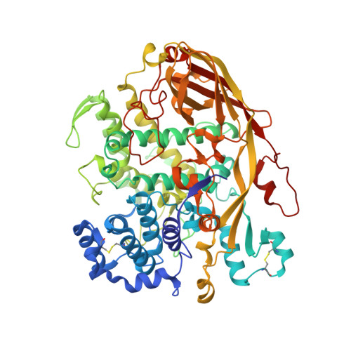
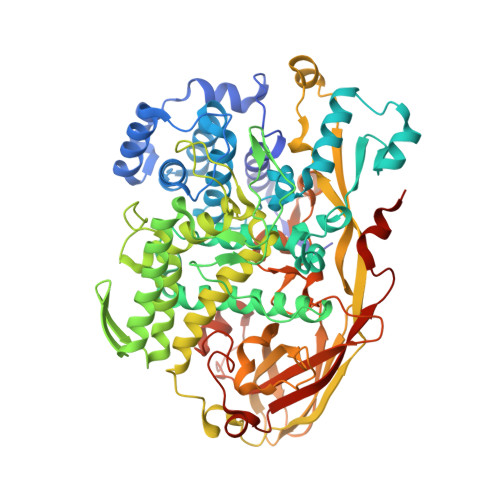
    The first crystal structure of a complex formed by two storage proteins, SP2 and SP3, isolated from their natural source, mulberry silkworm (Bombyx mori L.) haemolymph, has been determined. The structure was solved by molecular replacement using arylphorin, a protein rich in aromatic amino-acid residues, from oak silkworm as the initial model. The quality of the electron-density maps obtained from the X-ray diffraction experiment allowed the authors to detect that the investigated crystal structure was composed of two different arylphorins: SP2 and SP3. This discovery was confirmed by N-terminal sequencing. SP2 has been extensively studied previously, whereas only a few reports on SP3 are available. However, to date no structural studies have been reported for these proteins. These studies revealed that SP2 and SP3 exist in the silkworm body as a heterohexamer formed by one SP2 trimer and one SP3 trimer. The overall fold, consisting of three haemocyanin-like subdomains, of SP2 and SP3 is similar. Both proteins contain a conserved N-glycosylation motif in their structures.


  • Organizational Affiliation

    Center for Biocrystallographic Research, Institute of Bioorganic Chemistry, Polish Academy of Sciences, Noskowskiego 12/14, 61-704 Poznan, Poland.


Macromolecules
Find similar proteins by:  (by identity cutoff)  |  3D Structure
Entity ID: 1
MoleculeChains Sequence LengthOrganismDetailsImage
Silkworm storage protein680Bombyx moriMutation(s): 0 
UniProt
Find proteins for H9JHM9 (Bombyx mori)
Explore H9JHM9 
Go to UniProtKB:  H9JHM9
Entity Groups  
Sequence Clusters30% Identity50% Identity70% Identity90% Identity95% Identity100% Identity
UniProt GroupH9JHM9
Sequence Annotations
Expand
  • Reference Sequence
Find similar proteins by:  (by identity cutoff)  |  3D Structure
Entity ID: 2
MoleculeChains Sequence LengthOrganismDetailsImage
Arylphorin687Bombyx moriMutation(s): 0 
UniProt
Find proteins for Q1HPP4 (Bombyx mori)
Explore Q1HPP4 
Go to UniProtKB:  Q1HPP4
Entity Groups  
Sequence Clusters30% Identity50% Identity70% Identity90% Identity95% Identity100% Identity
UniProt GroupQ1HPP4
Sequence Annotations
Expand
  • Reference Sequence
Oligosaccharides

Help

Entity ID: 3
MoleculeChains Length2D Diagram Glycosylation3D Interactions
alpha-D-mannopyranose-(1-3)-[alpha-D-mannopyranose-(1-6)]beta-D-mannopyranose-(1-4)-2-acetamido-2-deoxy-beta-D-glucopyranose-(1-4)-2-acetamido-2-deoxy-beta-D-glucopyranose
C, D
5N-Glycosylation
Glycosylation Resources
GlyTouCan:  G22768VO
GlyCosmos:  G22768VO
GlyGen:  G22768VO
Small Molecules
Ligands 3 Unique
IDChains Name / Formula / InChI Key2D Diagram3D Interactions
SO4
Query on SO4

Download Ideal Coordinates CCD File 
E [auth A],
K [auth B]
SULFATE ION
O4 S
QAOWNCQODCNURD-UHFFFAOYSA-L
SCN
Query on SCN

Download Ideal Coordinates CCD File 
H [auth A],
I [auth A],
J [auth A],
M [auth B],
N [auth B]
THIOCYANATE ION
C N S
ZMZDMBWJUHKJPS-UHFFFAOYSA-M
NA
Query on NA

Download Ideal Coordinates CCD File 
F [auth A],
G [auth A],
L [auth B]
SODIUM ION
Na
FKNQFGJONOIPTF-UHFFFAOYSA-N
Experimental Data & Validation

Experimental Data

  • Method: X-RAY DIFFRACTION
  • Resolution: 2.90 Å
  • R-Value Free: 0.223 
  • R-Value Work: 0.166 
  • R-Value Observed: 0.167 
  • Space Group: P 63 2 2
Unit Cell:
Length ( Å )Angle ( ˚ )
a = 192.75α = 90
b = 192.75β = 90
c = 180.82γ = 120
Software Package:
Software NamePurpose
XDSdata scaling
PHASERphasing
REFMACrefinement
XDSdata reduction

Structure Validation

View Full Validation Report


Entry History 

Deposition Data

Revision History  (Full details and data files)

  • Version 1.0: 2013-12-04
    Type: Initial release
  • Version 1.1: 2014-09-24
    Changes: Database references
  • Version 2.0: 2020-07-29
    Type: Remediation
    Reason: Carbohydrate remediation
    Changes: Atomic model, Data collection, Derived calculations, Structure summary
  • Version 2.1: 2023-09-20
    Changes: Data collection, Database references, Refinement description, Structure summary