Structural Insights into the Mechanism for Recognizing Substrate of the Cytochrome P450 Enzyme TxtE.
Yu, F., Li, M.J., Xu, C.Y., Wang, Z.J., Zhou, H., Yang, M., Chen, Y.X., Tang, L., He, J.H.(2013) PLoS One 8: e81526-e81526
- PubMed: 24282603
- DOI: https://doi.org/10.1371/journal.pone.0081526
- Primary Citation of Related Structures:
4L36 - PubMed Abstract:
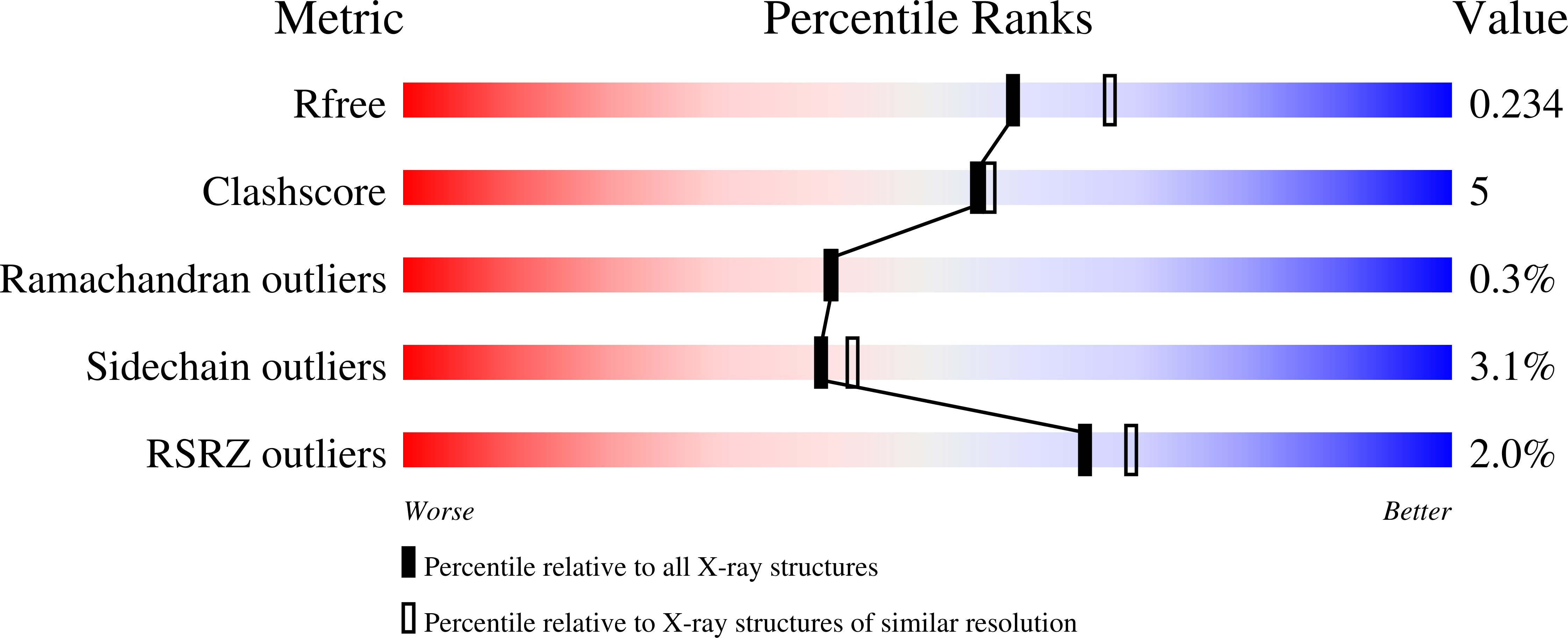
Thaxtomins, a family of phytotoxins produced by Streptomyces spp., can cause dramatic plant cell hypertrophy and seedling stunting. Thaxtomin A is the dominant form from Streptomyces scabies and has demonstrated herbicidal action. TxtE, a cytochrome P450 enzyme from Streptomyces scabies 87.22, catalyzes direct nitration of the indolyl moiety of L-tryptophan to L-4-nitrotryptophan using nitric oxide, dioxygen and NADPH. The crystal structure of TxtE was determined at 2.1 Å resolution and described in this work. A clearly defined substrate access channel is observed and can be classified as channel 2a, which is common in bacteria cytochrome P450 enzymes. A continuous hydrogen bond chain from the active site to the external solvent is observed. Compared with other cytochrome P450 enzymes, TxtE shows a unique proton transfer pathway which crosses the helix I distortion. Polar contacts of Arg59, Tyr89, Asn293, Thr296, and Glu394 with L-tryptophan are seen using molecular docking analysis, which are potentially important for substrate recognition and binding. After mutating Arg59, Asn293, Thr296 or Glu394 to leucine, the substrate binding ability of TxtE was lost or decreased significantly. Based on the docking and mutation results, a possible mechanism for substrate recognition and binding is proposed.
Organizational Affiliation:
Shanghai Institute of Applied Physics, Chinese Academy of Sciences, Shanghai, China.


