Structure of a translocation signal domain mediating conjugative transfer by type IV secretion systems.
Redzej, A., Ilangovan, A., Lang, S., Gruber, C.J., Topf, M., Zangger, K., Zechner, E.L., Waksman, G.(2013) Mol Microbiol 89: 324-333
- PubMed: 23710762
- DOI: https://doi.org/10.1111/mmi.12275
- Primary Citation of Related Structures:
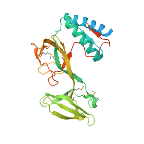
4L0J - PubMed Abstract:
Relaxases are proteins responsible for the transfer of plasmid and chromosomal DNA from one bacterium to another during conjugation. They covalently react with a specific phosphodiester bond within DNA origin of transfer sequences, forming a nucleo-protein complex which is subsequently recruited for transport by a plasmid-encoded type IV secretion system. In previous work we identified the targeting translocation signals presented by the conjugative relaxase TraI of plasmid R1. Here we report the structure of TraI translocation signal TSA. In contrast to known translocation signals we show that TSA is an independent folding unit and thus forms a bona fide structural domain. This domain can be further divided into three subdomains with striking structural homology with helicase subdomains of the SF1B family. We also show that TSA is part of a larger vestigial helicase domain which has lost its helicase activity but not its single-stranded DNA binding capability. Finally, we further delineate the binding site responsible for translocation activity of TSA by targeting single residues for mutations. Overall, this study provides the first evidence that translocation signals can be part of larger structural scaffolds, overlapping with translocation-independent activities.
Organizational Affiliation:
Institute of Structural and Molecular Biology, UCL and Birkbeck, Malet Street, London, WC1E 7HX, UK.