4L02

Crystal Structure of SphK1 with inhibitor


Experimental Data Snapshot

  • Method: X-RAY DIFFRACTION
  • Resolution: 2.75 Å
  • R-Value Free: 0.255 
  • R-Value Work: 0.208 
  • R-Value Observed: 0.210 

wwPDB Validation   3D Report Full Report


Ligand Structure Quality Assessment 


This is version 1.2 of the entry. See complete history


Literature

Structure guided design of a series of sphingosine kinase (SphK) inhibitors.

Gustin, D.J.Li, Y.Brown, M.L.Min, X.Schmitt, M.J.Wanska, M.Wang, X.Connors, R.Johnstone, S.Cardozo, M.Cheng, A.C.Jeffries, S.Franks, B.Li, S.Shen, S.Wong, M.Wesche, H.Xu, G.Carlson, T.J.Plant, M.Morgenstern, K.Rex, K.Schmitt, J.Coxon, A.Walker, N.Kayser, F.Wang, Z.

(2013) Bioorg Med Chem Lett 23: 4608-4616

  • DOI: https://doi.org/10.1016/j.bmcl.2013.06.030
  • Primary Citation of Related Structures:  
    4L02

  • PubMed Abstract: 

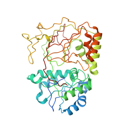
    Sphingosine-1-phosphate (S1P) signaling plays a vital role in mitogenesis, cell migration and angiogenesis. Sphingosine kinases (SphKs) catalyze a key step in sphingomyelin metabolism that leads to the production of S1P. There are two isoforms of SphK and observations made with SphK deficient mice show the two isoforms can compensate for each other's loss. Thus, inhibition of both isoforms is likely required to block SphK dependent angiogenesis. A structure based approach was used to design and synthesize a series of SphK inhibitors resulting in the identification of the first potent inhibitors of both isoforms of human SphK. Additionally, to our knowledge, this series of inhibitors contains the only sufficiently potent inhibitors of murine SphK1 with suitable physico-chemical properties to pharmacologically interrogate the role of SphK1 in rodent models and to reproduce the phenotype of SphK1 (-/-) mice.


  • Organizational Affiliation

    Department of Chemistry, Amgen Inc., 1120 Veterans Boulevard, South San Francisco, CA 94080, USA.


Macromolecules
Find similar proteins by:  (by identity cutoff)  |  3D Structure
Entity ID: 1
MoleculeChains Sequence LengthOrganismDetailsImage
Sphingosine kinase 1
A, B, C
384Homo sapiensMutation(s): 0 
Gene Names: Sphingosine kinase 1SPHKSPHK1SPK
EC: 2.7.1.91
UniProt & NIH Common Fund Data Resources
Find proteins for Q9NYA1 (Homo sapiens)
Explore Q9NYA1 
Go to UniProtKB:  Q9NYA1
PHAROS:  Q9NYA1
GTEx:  ENSG00000176170 
Entity Groups  
Sequence Clusters30% Identity50% Identity70% Identity90% Identity95% Identity100% Identity
UniProt GroupQ9NYA1
Sequence Annotations
Expand
  • Reference Sequence
Small Molecules
Ligands 1 Unique
IDChains Name / Formula / InChI Key2D Diagram3D Interactions
1V2
Query on 1V2

Download Ideal Coordinates CCD File 
D [auth A],
E [auth B],
F [auth C]
(2R,4S)-1-[2-(4-{[4-(3,4-dichlorophenyl)-1,3-thiazol-2-yl]amino}phenyl)ethyl]-2-(hydroxymethyl)piperidin-4-ol
C23 H25 Cl2 N3 O2 S
SQJKFWCRPARYPY-MOPGFXCFSA-N
Binding Affinity Annotations 
IDSourceBinding Affinity
1V2 PDBBind:  4L02 IC50: 20 (nM) from 1 assay(s)
BindingDB:  4L02 IC50: 20 (nM) from 1 assay(s)
Experimental Data & Validation

Experimental Data

  • Method: X-RAY DIFFRACTION
  • Resolution: 2.75 Å
  • R-Value Free: 0.255 
  • R-Value Work: 0.208 
  • R-Value Observed: 0.210 
  • Space Group: C 2 2 21
Unit Cell:
Length ( Å )Angle ( ˚ )
a = 102.575α = 90
b = 225.684β = 90
c = 106.411γ = 90
Software Package:
Software NamePurpose
BOSdata collection
PHASERphasing
REFMACrefinement
MOSFLMdata reduction
SCALAdata scaling

Structure Validation

View Full Validation Report


Ligand Structure Quality Assessment 


Entry History 

Deposition Data

Revision History  (Full details and data files)

  • Version 1.0: 2013-07-24
    Type: Initial release
  • Version 1.1: 2013-08-28
    Changes: Database references
  • Version 1.2: 2024-02-28
    Changes: Data collection, Database references, Derived calculations