4KYS

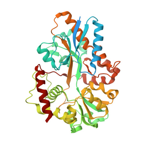
Clostridium botulinum thiaminase I in complex with thiamin


Experimental Data Snapshot

  • Method: X-RAY DIFFRACTION
  • Resolution: 2.18 Å
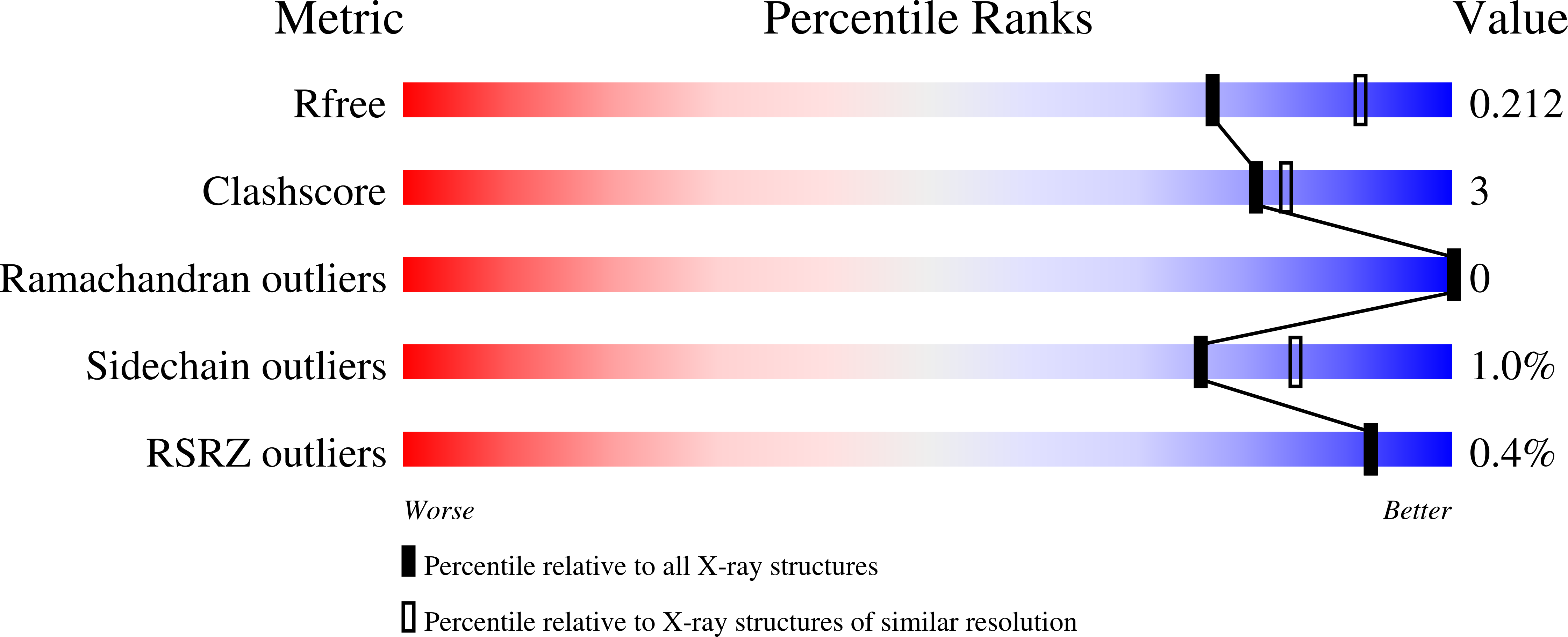
  • R-Value Free: 0.211 
  • R-Value Work: 0.156 
  • R-Value Observed: 0.159 

wwPDB Validation   3D Report Full Report


Ligand Structure Quality Assessment 


This is version 1.2 of the entry. See complete history


Literature

Structure of a Clostridium botulinum C143S Thiaminase I/Thiamin Complex Reveals Active Site Architecture.

Sikowitz, M.D.Shome, B.Zhang, Y.Begley, T.P.Ealick, S.E.

(2013) Biochemistry 52: 7830-7839

  • DOI: https://doi.org/10.1021/bi400841g
  • Primary Citation of Related Structures:  
    4KYS

  • PubMed Abstract: 

    Thiaminases are responsible for the degradation of thiamin and its metabolites. Two classes of thiaminases have been identified based on their three-dimensional structures and their requirements for a nucleophilic second substrate. Although the reactions of several thiaminases have been characterized, the physiological role of thiamin degradation is not fully understood. We have determined the three-dimensional X-ray structure of an inactive C143S mutant of Clostridium botulinum (Cb) thiaminase I with bound thiamin at 2.2 Å resolution. The C143S/thiamin complex provides atomic level details of the orientation of thiamin upon binding to Cb-thiaminase I and the identity of active site residues involved in substrate binding and catalysis. The specific roles of active site residues were probed by using site directed mutagenesis and kinetic analyses, leading to a detailed mechanism for Cb-thiaminase I. The structure of Cb-thiaminase I is also compared to the functionally similar but structurally distinct thiaminase II.


  • Organizational Affiliation

    Department of Chemistry and Chemical Biology, Cornell University , Ithaca, New York 14853, United States.


Macromolecules
Find similar proteins by:  (by identity cutoff)  |  3D Structure
Entity ID: 1
MoleculeChains Sequence LengthOrganismDetailsImage
Thiamine pyridinylase I
A, B
427Clostridium botulinum A str. ATCC 19397Mutation(s): 1 
Gene Names: CLB_0812
EC: 2.5.1.2
Entity Groups  
Sequence Clusters30% Identity50% Identity70% Identity90% Identity95% Identity100% Identity
Sequence Annotations
Expand
  • Reference Sequence
Small Molecules
Ligands 3 Unique
IDChains Name / Formula / InChI Key2D Diagram3D Interactions
VIB
Query on VIB

Download Ideal Coordinates CCD File 
C [auth A],
G [auth B]
3-(4-AMINO-2-METHYL-PYRIMIDIN-5-YLMETHYL)-5-(2-HYDROXY-ETHYL)-4-METHYL-THIAZOL-3-IUM
C12 H17 N4 O S
JZRWCGZRTZMZEH-UHFFFAOYSA-N
CIT
Query on CIT

Download Ideal Coordinates CCD File 
D [auth A],
H [auth B]
CITRIC ACID
C6 H8 O7
KRKNYBCHXYNGOX-UHFFFAOYSA-N
MG
Query on MG

Download Ideal Coordinates CCD File 
E [auth A],
F [auth A],
I [auth B],
J [auth B]
MAGNESIUM ION
Mg
JLVVSXFLKOJNIY-UHFFFAOYSA-N
Experimental Data & Validation

Experimental Data

  • Method: X-RAY DIFFRACTION
  • Resolution: 2.18 Å
  • R-Value Free: 0.211 
  • R-Value Work: 0.156 
  • R-Value Observed: 0.159 
  • Space Group: P 1 21 1
Unit Cell:
Length ( Å )Angle ( ˚ )
a = 63.504α = 90
b = 36.306β = 91.44
c = 156.813γ = 90
Software Package:
Software NamePurpose
ADSCdata collection
PHENIXmodel building
PHENIXrefinement
XDSdata reduction
SCALAdata scaling
PHENIXphasing

Structure Validation

View Full Validation Report


Ligand Structure Quality Assessment 


Entry History 

Deposition Data

Revision History  (Full details and data files)

  • Version 1.0: 2013-10-30
    Type: Initial release
  • Version 1.1: 2013-11-20
    Changes: Database references
  • Version 1.2: 2024-02-28
    Changes: Data collection, Database references, Derived calculations, Structure summary