4KQ2

Glucose1,2cyclic phosphate bound activated state of Yeast Glycogen Synthase


Experimental Data Snapshot

  • Method: X-RAY DIFFRACTION
  • Resolution: 2.95 Å
  • R-Value Free: 0.271 
  • R-Value Work: 0.224 
  • R-Value Observed: 0.227 

wwPDB Validation   3D Report Full Report


Ligand Structure Quality Assessment 


This is version 1.5 of the entry. See complete history


Literature

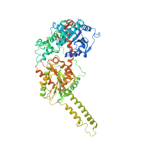
Structural basis for 2'-phosphate incorporation into glycogen by glycogen synthase.

Chikwana, V.M.Khanna, M.Baskaran, S.Tagliabracci, V.S.Contreras, C.J.Depaoli-Roach, A.Roach, P.J.Hurley, T.D.

(2013) Proc Natl Acad Sci U S A 110: 20976-20981

  • DOI: https://doi.org/10.1073/pnas.1310106111
  • Primary Citation of Related Structures:  
    4KQ1, 4KQ2, 4KQM

  • PubMed Abstract: 

    Glycogen is a glucose polymer that contains minor amounts of covalently attached phosphate. Hyperphosphorylation is deleterious to glycogen structure and can lead to Lafora disease. Recently, it was demonstrated that glycogen synthase catalyzes glucose-phosphate transfer in addition to its characteristic glucose transfer reaction. Glucose-1,2-cyclic-phosphate (GCP) was proposed to be formed from UDP-Glc breakdown and subsequently transferred, thus providing a source of phosphate found in glycogen. To gain further insight into the molecular basis for glucose-phosphate transfer, two structures of yeast glycogen synthase were determined; a 3.0-Å resolution structure of the complex with UMP/GCP and a 2.8-Å resolution structure of the complex with UDP/glucose. Structural superposition of the complexes revealed that the bound ligands and most active site residues are positioned similarly, consistent with the use of a common transfer mechanism for both reactions. The N-terminal domain of the UDP-glucose complex was found to be 13.3° more closed compared with a UDP complex. However, the UMP · GCP complex was 4.8° less closed than the glucose complex, which may explain the low efficiency of GCP transfer. Modeling of either α- or β-glucose or a mixture of both anomers can account for the observed electron density of the UDP-glucose complex. NMR studies of UDP-Glc hydrolysis by yeast glycogen synthase were used to verify the stereochemistry of the product, and they also showed synchronous GCP accumulation. The similarities in the active sites of glycogen synthase and glycogen phosphorylase support the idea of a common catalytic mechanism in GT-B enzymes independent of the specific reaction catalyzed.


  • Organizational Affiliation

    Department of Biochemistry and Molecular Biology, Indiana University School of Medicine, Indianapolis, IN 46202.


Macromolecules
Find similar proteins by:  (by identity cutoff)  |  3D Structure
Entity ID: 1
MoleculeChains Sequence LengthOrganismDetailsImage
Gsy2p
A, B, C, D
724Saccharomyces cerevisiae FostersOMutation(s): 2 
Gene Names: FOSTERSO_3265GSY2 YLR258W L8479.8
UniProt
Find proteins for P27472 (Saccharomyces cerevisiae (strain ATCC 204508 / S288c))
Explore P27472 
Go to UniProtKB:  P27472
Entity Groups  
Sequence Clusters30% Identity50% Identity70% Identity90% Identity95% Identity100% Identity
UniProt GroupP27472
Sequence Annotations
Expand
  • Reference Sequence
Small Molecules
Ligands 5 Unique
IDChains Name / Formula / InChI Key2D Diagram3D Interactions
U5P
Query on U5P

Download Ideal Coordinates CCD File 
E [auth A],
J [auth B],
P [auth C],
T [auth D]
URIDINE-5'-MONOPHOSPHATE
C9 H13 N2 O9 P
DJJCXFVJDGTHFX-XVFCMESISA-N
G6P
Query on G6P

Download Ideal Coordinates CCD File 
F [auth A],
K [auth B],
Q [auth C],
U [auth D]
6-O-phosphono-alpha-D-glucopyranose
C6 H13 O9 P
NBSCHQHZLSJFNQ-DVKNGEFBSA-N
1S3
Query on 1S3

Download Ideal Coordinates CCD File 
O [auth C](2R,3aR,5R,6S,7S,7aR)-5-(hydroxymethyl)tetrahydro-3aH-[1,3,2]dioxaphospholo[4,5-b]pyran-2,6,7-triol 2-oxide
C6 H11 O8 P
PUJJEEQZLDDQPG-VFUOTHLCSA-N
BA
Query on BA

Download Ideal Coordinates CCD File 
H [auth A]
I [auth A]
M [auth B]
N [auth B]
S [auth C]
H [auth A],
I [auth A],
M [auth B],
N [auth B],
S [auth C],
W [auth D],
X [auth D]
BARIUM ION
Ba
XDFCIPNJCBUZJN-UHFFFAOYSA-N
PEG
Query on PEG

Download Ideal Coordinates CCD File 
G [auth A],
L [auth B],
R [auth C],
V [auth D]
DI(HYDROXYETHYL)ETHER
C4 H10 O3
MTHSVFCYNBDYFN-UHFFFAOYSA-N
Experimental Data & Validation

Experimental Data

  • Method: X-RAY DIFFRACTION
  • Resolution: 2.95 Å
  • R-Value Free: 0.271 
  • R-Value Work: 0.224 
  • R-Value Observed: 0.227 
  • Space Group: I 2 2 2
Unit Cell:
Length ( Å )Angle ( ˚ )
a = 193.49α = 90
b = 203.978β = 90
c = 206.307γ = 90
Software Package:
Software NamePurpose
DENZOdata reduction
SCALEPACKdata scaling
REFMACrefinement
PDB_EXTRACTdata extraction
HKL-3000data collection
HKL-3000data reduction
MOLREPphasing

Structure Validation

View Full Validation Report


Ligand Structure Quality Assessment 


Entry History 

Deposition Data

Revision History  (Full details and data files)

  • Version 1.0: 2013-12-11
    Type: Initial release
  • Version 1.1: 2014-02-05
    Changes: Database references
  • Version 1.2: 2014-11-12
    Changes: Structure summary
  • Version 1.3: 2017-11-15
    Changes: Refinement description
  • Version 1.4: 2020-07-29
    Type: Remediation
    Reason: Carbohydrate remediation
    Changes: Data collection, Database references, Derived calculations, Structure summary
  • Version 1.5: 2023-09-20
    Changes: Data collection, Database references, Refinement description, Structure summary