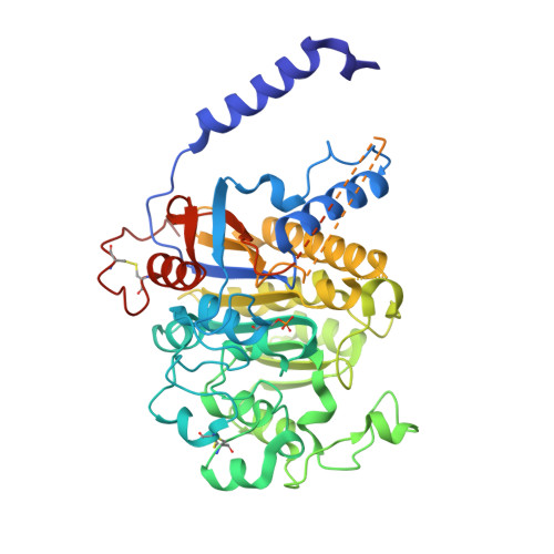
Crystal structure of rat intestinal alkaline phosphatase - Role of crown domain in mammalian alkaline phosphatases.
Ghosh, K., Mazumder Tagore, D., Anumula, R., Lakshmaiah, B., Kumar, P.P., Singaram, S., Matan, T., Kallipatti, S., Selvam, S., Krishnamurthy, P., Ramarao, M.(2013) J Struct Biol 184: 182-192
- PubMed: 24076154
- DOI: https://doi.org/10.1016/j.jsb.2013.09.017
- Primary Citation of Related Structures:
4KJD, 4KJG - PubMed Abstract:
Intestinal alkaline phosphatases (IAPs) are involved in the cleavage of phosphate prodrugs to liberate the drug for absorption in the intestine. To facilitate in vitro characterization of phosphate prodrugs, we have cloned, expressed, purified and characterized IAPs from rat and cynomolgus monkey (rIAP and cIAP respectively) which are important pre-clinical species for drug metabolism studies. The recombinant rat and monkey enzymes expressed in Sf9 insect cells (IAP-Ic) were found to be glycosylated and active. Expression of rat IAP in Escherichia coli (rIAP-Ec) led to ~200-fold loss of activity that was partially recovered by the addition of external Zn(2+) and Mg(2+) ions. Crystal structures of rIAP-Ec and rIAP-Ic were determined and they provide rationale for the discrepancy in enzyme activities. Rat IAP-Ic retains its activity in presence of both Zn(2+) and Mg(2+) whereas activity of most other alkaline phosphatases (APs) including the cIAP was strongly inhibited by excess Zn(2+). Based on our crystal structure, we hypothesized the residue Q317 in rIAP, present within 7 Å of the Mg(2+) at M3, to be important for this difference in activity. The Q317H rIAP and H317Q cIAP mutants showed reversal in effect of Zn(2+), corroborating the hypothesis. Further analysis of the two structures indicated a close linkage between glycosylation and crown domain stability. A triple mutant of rIAP, where all the three putative N-linked glycosylation sites were mutated showed thermal instability and reduced activity.
Organizational Affiliation:
Applied Biotechnology, Biocon Bristol-Myers Squibb Research and Development Center, Syngene International Ltd., Biocon Park, Bommasandra IV Phase, Jigani Link Road, Bangalore 560099, India. Electronic address: Kaushik.Ghosh@syngeneintl.com.