4KFQ

Crystal structure of the NMDA receptor GluN1 ligand binding domain in complex with 1-thioxo-1,2-dihydro-[1,2,4]triazolo[4,3-a]quinoxalin-4(5H)-one


Experimental Data Snapshot

  • Method: X-RAY DIFFRACTION
  • Resolution: 2.20 Å
  • R-Value Free: 0.232 
  • R-Value Work: 0.174 
  • R-Value Observed: 0.177 

wwPDB Validation   3D Report Full Report


Ligand Structure Quality Assessment 


This is version 1.3 of the entry. See complete history


Literature

Crystal structure and pharmacological characterization of a novel N-methyl-D-aspartate (NMDA) receptor antagonist at the GluN1 glycine binding site.

Kvist, T.Steffensen, T.B.Greenwood, J.R.Mehrzad Tabrizi, F.Hansen, K.B.Gajhede, M.Pickering, D.S.Traynelis, S.F.Kastrup, J.S.Brauner-Osborne, H.

(2013) J Biol Chem 288: 33124-33135

  • DOI: https://doi.org/10.1074/jbc.M113.480210
  • Primary Citation of Related Structures:  
    4KFQ

  • PubMed Abstract: 

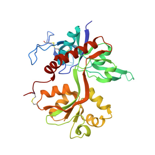
    NMDA receptors are ligand-gated ion channels that mediate excitatory neurotransmission in the brain. They are tetrameric complexes composed of glycine-binding GluN1 and GluN3 subunits together with glutamate-binding GluN2 subunits. Subunit-selective antagonists that discriminate between the glycine sites of GluN1 and GluN3 subunits would be valuable pharmacological tools for studies on the function and physiological roles of NMDA receptor subtypes. In a virtual screening for antagonists that exploit differences in the orthosteric binding site of GluN1 and GluN3 subunits, we identified a novel glycine site antagonist, 1-thioxo-1,2-dihydro-[1,2,4]triazolo[4,3-a]quinoxalin-4(5H)-one (TK40). Here, we show by Schild analysis that TK40 is a potent competitive antagonist with Kb values of 21-63 nM at the GluN1 glycine-binding site of the four recombinant GluN1/N2A-D receptors. In addition, TK40 displayed >100-fold selectivity for GluN1/N2 NMDA receptors over GluN3A- and GluN3B-containing NMDA receptors and no appreciable effects at AMPA receptors. Binding experiments on rat brain membranes and the purified GluN1 ligand-binding domain using glycine site GluN1 radioligands further confirmed the competitive interaction and high potency. To delineate the binding mechanism, we have solved the crystal structure of the GluN1 ligand-binding domain in complex with TK40 and show that TK40 binds to the orthosteric binding site of the GluN1 subunit with a binding mode that was also predicted by virtual screening. Furthermore, the structure reveals that the imino acetamido group of TK40 acts as an α-amino acid bioisostere, which could be of importance in bioisosteric replacement strategies for future ligand design.


  • Organizational Affiliation

    From the Department of Drug Design and Pharmacology, Faculty of Health and Medical Sciences, University of Copenhagen, Universitetsparken 2, DK-2100 Copenhagen, Denmark.


Macromolecules
Find similar proteins by:  (by identity cutoff)  |  3D Structure
Entity ID: 1
MoleculeChains Sequence LengthOrganismDetailsImage
Glutamate receptor ionotropic, NMDA 1
A, B
292Rattus norvegicusMutation(s): 0 
Gene Names: Grin1GRIN1 OR NMDAR1Nmdar1
UniProt
Find proteins for P35439 (Rattus norvegicus)
Explore P35439 
Go to UniProtKB:  P35439
Entity Groups  
Sequence Clusters30% Identity50% Identity70% Identity90% Identity95% Identity100% Identity
UniProt GroupP35439
Sequence Annotations
Expand
  • Reference Sequence
Small Molecules
Ligands 3 Unique
IDChains Name / Formula / InChI Key2D Diagram3D Interactions
KFQ
Query on KFQ

Download Ideal Coordinates CCD File 
C [auth A],
Q [auth B]
1-sulfanyl[1,2,4]triazolo[4,3-a]quinoxalin-4(5H)-one
C9 H6 N4 O S
WDUVQRMZWGBWJS-UHFFFAOYSA-N
SO4
Query on SO4

Download Ideal Coordinates CCD File 
AA [auth B]
BA [auth B]
CA [auth B]
DA [auth B]
M [auth A]
AA [auth B],
BA [auth B],
CA [auth B],
DA [auth B],
M [auth A],
N [auth A],
O [auth A],
P [auth A]
SULFATE ION
O4 S
QAOWNCQODCNURD-UHFFFAOYSA-L
GOL
Query on GOL

Download Ideal Coordinates CCD File 
D [auth A]
E [auth A]
F [auth A]
G [auth A]
H [auth A]
D [auth A],
E [auth A],
F [auth A],
G [auth A],
H [auth A],
I [auth A],
J [auth A],
K [auth A],
L [auth A],
R [auth B],
S [auth B],
T [auth B],
U [auth B],
V [auth B],
W [auth B],
X [auth B],
Y [auth B],
Z [auth B]
GLYCEROL
C3 H8 O3
PEDCQBHIVMGVHV-UHFFFAOYSA-N
Experimental Data & Validation

Experimental Data

  • Method: X-RAY DIFFRACTION
  • Resolution: 2.20 Å
  • R-Value Free: 0.232 
  • R-Value Work: 0.174 
  • R-Value Observed: 0.177 
  • Space Group: P 1 21 1
Unit Cell:
Length ( Å )Angle ( ˚ )
a = 43.95α = 90
b = 78.59β = 94.21
c = 109.63γ = 90
Software Package:
Software NamePurpose
MxCuBEdata collection
PHASERphasing
PHENIXrefinement
MOSFLMdata reduction
SCALAdata scaling

Structure Validation

View Full Validation Report


Ligand Structure Quality Assessment 


Entry History 

Deposition Data

Revision History  (Full details and data files)

  • Version 1.0: 2013-10-09
    Type: Initial release
  • Version 1.1: 2015-07-01
    Changes: Database references, Structure summary
  • Version 1.2: 2017-08-23
    Changes: Refinement description, Source and taxonomy
  • Version 1.3: 2023-09-20
    Changes: Data collection, Database references, Derived calculations, Refinement description