4KE2

Crystal structure of the hyperactive Type I antifreeze from winter flounder


Experimental Data Snapshot

  • Method: X-RAY DIFFRACTION
  • Resolution: 1.80 Å
  • R-Value Free: 0.253 
  • R-Value Work: 0.196 
  • R-Value Observed: 0.199 

wwPDB Validation   3D Report Full Report


This is version 1.2 of the entry. See complete history


Literature

An antifreeze protein folds with an interior network of more than 400 semi-clathrate waters.

Sun, T.Lin, F.H.Campbell, R.L.Allingham, J.S.Davies, P.L.

(2014) Science 343: 795-798

  • DOI: https://doi.org/10.1126/science.1247407
  • Primary Citation of Related Structures:  
    4KE2

  • PubMed Abstract: 

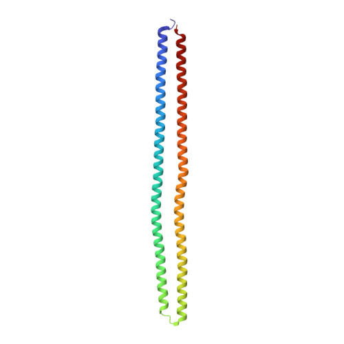
    When polypeptide chains fold into a protein, hydrophobic groups are compacted in the center with exclusion of water. We report the crystal structure of an alanine-rich antifreeze protein that retains ~400 waters in its core. The putative ice-binding residues of this dimeric, four-helix bundle protein point inwards and coordinate the interior waters into two intersecting polypentagonal networks. The bundle makes minimal protein contacts between helices, but is stabilized by anchoring to the semi-clathrate water monolayers through backbone carbonyl groups in the protein interior. The ordered waters extend outwards to the protein surface and likely are involved in ice binding. This protein fold supports both the anchored-clathrate water mechanism of antifreeze protein adsorption to ice and the water-expulsion mechanism of protein folding.


  • Organizational Affiliation

    Department of Biomedical and Molecular Sciences, Queen's University, Kingston, ON K7L 3N6, Canada.


Macromolecules
Find similar proteins by:  (by identity cutoff)  |  3D Structure
Entity ID: 1
MoleculeChains Sequence LengthOrganismDetailsImage
Type I hyperactive antifreeze protein
A, B, C
196Pseudopleuronectes americanusMutation(s): 0 
UniProt
Find proteins for B1P0S1 (Pseudopleuronectes americanus)
Explore B1P0S1 
Go to UniProtKB:  B1P0S1
Entity Groups  
Sequence Clusters30% Identity50% Identity70% Identity90% Identity95% Identity100% Identity
UniProt GroupB1P0S1
Sequence Annotations
Expand
  • Reference Sequence
Experimental Data & Validation

Experimental Data

  • Method: X-RAY DIFFRACTION
  • Resolution: 1.80 Å
  • R-Value Free: 0.253 
  • R-Value Work: 0.196 
  • R-Value Observed: 0.199 
  • Space Group: C 1 2 1
Unit Cell:
Length ( Å )Angle ( ˚ )
a = 95.82α = 90
b = 48.14β = 91.87
c = 182.54γ = 90
Software Package:
Software NamePurpose
XSCALEdata scaling
SHARPphasing
PHENIXrefinement
PDB_EXTRACTdata extraction
XDSdata reduction
autoSHARPphasing

Structure Validation

View Full Validation Report


Entry History 

Deposition Data

Revision History  (Full details and data files)

  • Version 1.0: 2014-02-26
    Type: Initial release
  • Version 1.1: 2014-09-24
    Changes: Database references
  • Version 1.2: 2024-02-28
    Changes: Data collection, Database references