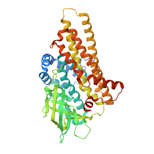
4KCF

X-ray Structure of a KijD3 in Complex with FMN and dTDP-3-amino-2,3,6-trideoxy-4-keto-3-methyl-D-glucose


Experimental Data Snapshot

  • Method: X-RAY DIFFRACTION
  • Resolution: 2.10 Å
  • R-Value Free: 0.225 
  • R-Value Work: 0.175 
  • R-Value Observed: 0.177 

wwPDB Validation   3D Report Full Report


Ligand Structure Quality Assessment 


This is version 1.3 of the entry. See complete history


Literature

Active site architecture of a sugar N-oxygenase.

Thoden, J.B.Branch, M.C.Zimmer, A.L.Bruender, N.A.Holden, H.M.

(2013) Biochemistry 52: 3191-3193

  • DOI: https://doi.org/10.1021/bi400407x
  • Primary Citation of Related Structures:  
    4KCF

  • PubMed Abstract: 

    KijD3 is a flavin-dependent N-oxygenase implicated in the formation of the nitro-containing sugar d-kijanose, found attached to the antibiotic kijanimicin. For this investigation, the structure of KijD3 in complex with FMN and its dTDP-sugar substrate was solved to 2.1 Å resolution. In contrast to the apoenzyme structure, the C-terminus of the protein becomes ordered and projects into the active site cleft [Bruender, N. A., Thoden, J. B., and Holden, H. M. (2010) Biochemistry 49, 3517-3524]. The amino group of the dTDP-aminosugar that is oxidized is located 4.9 Å from C4a of the flavin ring. The model provides a molecular basis for understanding the manner in which KijD3 catalyzes its unusual chemical transformation.


  • Organizational Affiliation

    Department of Biochemistry, University of Wisconsin , Madison, Wisconsin 53706, United States.


Macromolecules
Find similar proteins by:  (by identity cutoff)  |  3D Structure
Entity ID: 1
MoleculeChains Sequence LengthOrganismDetailsImage
FAD-dependent oxidoreductase454Actinomadura kijaniataMutation(s): 0 
Gene Names: KijD3
UniProt
Find proteins for B3TMR1 (Actinomadura kijaniata)
Explore B3TMR1 
Go to UniProtKB:  B3TMR1
Entity Groups  
Sequence Clusters30% Identity50% Identity70% Identity90% Identity95% Identity100% Identity
UniProt GroupB3TMR1
Sequence Annotations
Expand
  • Reference Sequence
Small Molecules
Ligands 2 Unique
IDChains Name / Formula / InChI Key2D Diagram3D Interactions
AKM
Query on AKM

Download Ideal Coordinates CCD File 
B [auth A][(2R,4S,6R)-4-azanyl-4,6-dimethyl-5,5-bis(oxidanyl)oxan-2-yl] [[(2R,3S,5R)-5-[5-methyl-2,4-bis(oxidanylidene)pyrimidin-1-yl]-3-oxidanyl-oxolan-2-yl]methoxy-oxidanyl-phosphoryl] hydrogen phosphate
C17 H29 N3 O14 P2
GACNZVKWDHTCBB-HHMBNNFFSA-N
FMN
Query on FMN

Download Ideal Coordinates CCD File 
C [auth A]FLAVIN MONONUCLEOTIDE
C17 H21 N4 O9 P
FVTCRASFADXXNN-SCRDCRAPSA-N
Experimental Data & Validation

Experimental Data

  • Method: X-RAY DIFFRACTION
  • Resolution: 2.10 Å
  • R-Value Free: 0.225 
  • R-Value Work: 0.175 
  • R-Value Observed: 0.177 
  • Space Group: I 2 2 2
Unit Cell:
Length ( Å )Angle ( ˚ )
a = 70.828α = 90
b = 81.297β = 90
c = 152.628γ = 90
Software Package:
Software NamePurpose
HKL-3000data collection
PHASERphasing
REFMACrefinement
HKL-3000data reduction
HKL-3000data scaling

Structure Validation

View Full Validation Report


Ligand Structure Quality Assessment 


Entry History 

Deposition Data

Revision History  (Full details and data files)

  • Version 1.0: 2013-05-22
    Type: Initial release
  • Version 1.1: 2013-05-29
    Changes: Atomic model, Non-polymer description
  • Version 1.2: 2013-12-18
    Changes: Database references
  • Version 1.3: 2023-09-20
    Changes: Data collection, Database references, Derived calculations, Refinement description