4KC2

Structure of the blood group glycosyltransferase AAglyB in complex with a pyridine inhibitor as a neutral pyrophosphate surrogate


Experimental Data Snapshot

  • Method: X-RAY DIFFRACTION
  • Resolution: 1.70 Å
  • R-Value Free: 0.205 
  • R-Value Work: 0.176 
  • R-Value Observed: 0.177 

wwPDB Validation   3D Report Full Report


Ligand Structure Quality Assessment 


This is version 1.3 of the entry. See complete history


Literature

Design of glycosyltransferase inhibitors: pyridine as a pyrophosphate surrogate.

Wang, S.Cuesta-Seijo, J.A.Lafont, D.Palcic, M.M.Vidal, S.

(2013) Chemistry 19: 15346-15357

  • DOI: https://doi.org/10.1002/chem.201301871
  • Primary Citation of Related Structures:  
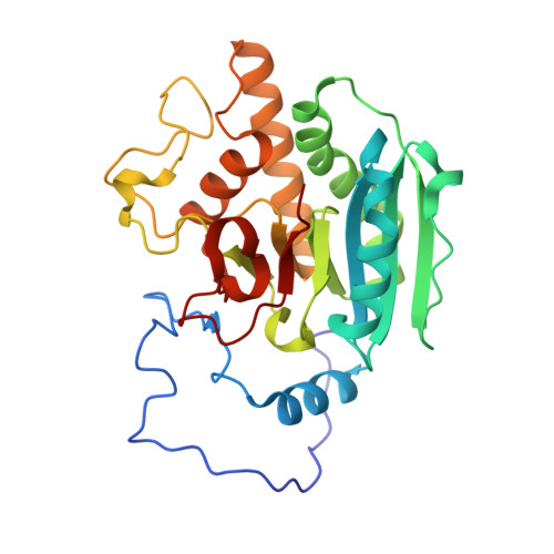
    4KC1, 4KC2, 4KC4

  • PubMed Abstract: 

    A series of ten glycosyltransferase inhibitors has been designed and synthesized by using pyridine as a pyrophosphate surrogate. The series was prepared by conjugation of carbohydrate, pyridine, and nucleoside building blocks by using a combination of glycosylation, the Staudinger-Vilarrasa amide-bond formation, and azide-alkyne click chemistry. The compounds were evaluated as inhibitors of five metal-dependent galactosyltransferases. Crystallographic analyses of three inhibitors complexed in the active site of one of the enzymes confirmed that the pyridine moiety chelates the Mn(2+) ion causing a slight displacement (2 Å) from its original position. The carbohydrate head group occupies a different position than in the natural uridine diphosphate (UDP)-Gal substrate with little interaction with the enzyme.


  • Organizational Affiliation

    Institut de Chimie et Biochimie Moléculaires et Supramoléculaires, Laboratoire de Chimie Organique 2, Glycochimie, UMR 5246, CNRS and Université Claude Bernard Lyon 1, 43 Boulevard du 11 Novembre 1918, 6922 Villeurbanne (France), Fax: (+33) 472-448-109.


Macromolecules
Find similar proteins by:  (by identity cutoff)  |  3D Structure
Entity ID: 1
MoleculeChains Sequence LengthOrganismDetailsImage
Fucosylglycoprotein alpha-N-acetylgalactosaminyltransferase soluble form290Homo sapiensMutation(s): 0 
Gene Names: ABO
EC: 2.4.1.40 (PDB Primary Data), 2.4.1.37 (PDB Primary Data)
UniProt & NIH Common Fund Data Resources
Find proteins for P16442 (Homo sapiens)
Explore P16442 
Go to UniProtKB:  P16442
PHAROS:  P16442
Entity Groups  
Sequence Clusters30% Identity50% Identity70% Identity90% Identity95% Identity100% Identity
UniProt GroupP16442
Sequence Annotations
Expand
  • Reference Sequence
Small Molecules
Ligands 3 Unique
IDChains Name / Formula / InChI Key2D Diagram3D Interactions
WS2
Query on WS2

Download Ideal Coordinates CCD File 
D [auth A]6-(1-beta-D-Galactopyranosyloxymethyl)-N-(5'-deoxyluridine-5'-yl)picolinamide
C22 H28 N4 O12
KPOTWZYLDCHCIG-GBMKPDNQSA-N
BHE
Query on BHE

Download Ideal Coordinates CCD File 
C [auth A]octyl 2-O-(6-deoxy-alpha-L-galactopyranosyl)-beta-D-galactopyranoside
C20 H38 O10
GTTDTLMUWQMDNA-ARNYJBIMSA-N
MN
Query on MN

Download Ideal Coordinates CCD File 
B [auth A]MANGANESE (II) ION
Mn
WAEMQWOKJMHJLA-UHFFFAOYSA-N
Experimental Data & Validation

Experimental Data

  • Method: X-RAY DIFFRACTION
  • Resolution: 1.70 Å
  • R-Value Free: 0.205 
  • R-Value Work: 0.176 
  • R-Value Observed: 0.177 
  • Space Group: C 2 2 21
Unit Cell:
Length ( Å )Angle ( ˚ )
a = 52.88α = 90
b = 149.9β = 90
c = 80.03γ = 90
Software Package:
Software NamePurpose
XSCALEdata scaling
REFMACrefinement
PDB_EXTRACTdata extraction
MAR345dtbdata collection
XDSdata reduction

Structure Validation

View Full Validation Report


Ligand Structure Quality Assessment 


Entry History 

Deposition Data

Revision History  (Full details and data files)

  • Version 1.0: 2013-09-11
    Type: Initial release
  • Version 1.1: 2015-03-04
    Changes: Database references
  • Version 1.2: 2017-11-15
    Changes: Advisory, Database references, Refinement description
  • Version 1.3: 2023-09-20
    Changes: Data collection, Database references, Derived calculations, Refinement description