4KBY

mSTING/c-di-GMP


Experimental Data Snapshot

  • Method: X-RAY DIFFRACTION
  • Resolution: 2.36 Å
  • R-Value Free: 0.265 
  • R-Value Work: 0.181 
  • R-Value Observed: 0.190 

wwPDB Validation   3D Report Full Report


Ligand Structure Quality Assessment 


This is version 1.1 of the entry. See complete history


Literature

Novel c-di-GMP recognition modes of the mouse innate immune adaptor protein STING

Chin, K.H.Tu, Z.L.Su, Y.C.Yu, Y.J.Chen, H.C.Lo, Y.C.Chen, C.P.Barber, G.N.Chuah, M.L.Liang, Z.X.Chou, S.H.

(2013) Acta Crystallogr D Biol Crystallogr 69: 352-366

  • DOI: https://doi.org/10.1107/S0907444912047269
  • Primary Citation of Related Structures:  
    4KBY, 4KC0

  • PubMed Abstract: 

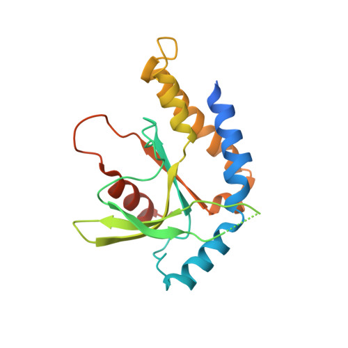
    The mammalian ER protein STING (stimulator of interferon genes; also known as MITA, ERIS, MPYS or TMEM173) is an adaptor protein that links the detection of cytosolic dsDNA to the activation of TANK-binding kinase 1 (TBK1) and its downstream transcription factor interferon regulatory factor 3 (IFN3). Recently, STING itself has been found to be the direct receptor of bacterial c-di-GMP, and crystal structures of several human STING C-terminal domain (STING-CTD) dimers in the apo form or in complex with c-di-GMP have been published. Here, a novel set of structures of mouse STING-CTD (mSTING(137-344)) in apo and complex forms determined from crystals obtained under different crystallization conditions are reported. These novel closed-form structures exhibited considerable differences from previously reported open-form human STING-CTD structures. The novel mSTING structures feature extensive interactions between the two monomers, a unique asymmetric c-di-GMP molecule with one guanine base in an unusual syn conformation that is well accommodated in the dimeric interface with many direct specific interactions and two unexpected equivalent secondary peripheral c-di-GMP binding sites. Replacement of the amino acids crucial for specific c-di-GMP binding in mSTING significantly changes the ITC titration profiles and reduces the IFN-β reporter luciferase activity. Taken together, these results reveal a more stable c-di-GMP binding mode of STING proteins that could serve as a template for rational drug design to stimulate interferon production by mammalian cells.


  • Organizational Affiliation

    Agricultural Biotechnology Center, National Chung Hsing University, Taichung 40227, Taiwan.


Macromolecules
Find similar proteins by:  (by identity cutoff)  |  3D Structure
Entity ID: 1
MoleculeChains Sequence LengthOrganismDetailsImage
Stimulator of interferon genes protein
A, B
207Mus musculusMutation(s): 0 
Gene Names: Tmem173Eris MitaMpysSting
UniProt & NIH Common Fund Data Resources
Find proteins for Q3TBT3 (Mus musculus)
Explore Q3TBT3 
Go to UniProtKB:  Q3TBT3
IMPC:  MGI:1919762
Entity Groups  
Sequence Clusters30% Identity50% Identity70% Identity90% Identity95% Identity100% Identity
UniProt GroupQ3TBT3
Sequence Annotations
Expand
  • Reference Sequence
Small Molecules
Ligands 1 Unique
IDChains Name / Formula / InChI Key2D Diagram3D Interactions
C2E
Query on C2E

Download Ideal Coordinates CCD File 
C [auth A]9,9'-[(2R,3R,3aS,5S,7aR,9R,10R,10aS,12S,14aR)-3,5,10,12-tetrahydroxy-5,12-dioxidooctahydro-2H,7H-difuro[3,2-d:3',2'-j][1,3,7,9,2,8]tetraoxadiphosphacyclododecine-2,9-diyl]bis(2-amino-1,9-dihydro-6H-purin-6-one)
C20 H24 N10 O14 P2
PKFDLKSEZWEFGL-MHARETSRSA-N
Binding Affinity Annotations 
IDSourceBinding Affinity
C2E PDBBind:  4KBY Kd: 186 (nM) from 1 assay(s)
Binding MOAD:  4KBY Ka: 5.39e+6 (M^-1) from 1 assay(s)
Experimental Data & Validation

Experimental Data

  • Method: X-RAY DIFFRACTION
  • Resolution: 2.36 Å
  • R-Value Free: 0.265 
  • R-Value Work: 0.181 
  • R-Value Observed: 0.190 
  • Space Group: P 31
Unit Cell:
Length ( Å )Angle ( ˚ )
a = 79.058α = 90
b = 79.058β = 90
c = 49.693γ = 120
Software Package:
Software NamePurpose
HKL-2000data collection
CNSrefinement
PHENIXrefinement
HKL-2000data reduction
HKL-2000data scaling
CNSphasing

Structure Validation

View Full Validation Report


Ligand Structure Quality Assessment 


Entry History 

Deposition Data

Revision History  (Full details and data files)

  • Version 1.0: 2013-05-29
    Type: Initial release
  • Version 1.1: 2024-03-20
    Changes: Data collection, Database references, Derived calculations, Structure summary