4KAX

Crystal structure of the Grp1 PH domain in complex with Arf6-GTP


Experimental Data Snapshot

  • Method: X-RAY DIFFRACTION
  • Resolution: 1.85 Å
  • R-Value Free: 0.235 
  • R-Value Work: 0.200 
  • R-Value Observed: 0.202 

wwPDB Validation   3D Report Full Report


Ligand Structure Quality Assessment 


This is version 1.4 of the entry. See complete history


Literature

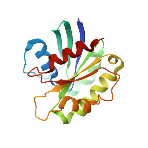
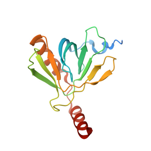
Structural basis for membrane recruitment and allosteric activation of cytohesin family Arf GTPase exchange factors.

Malaby, A.W.van den Berg, B.Lambright, D.G.

(2013) Proc Natl Acad Sci U S A 110: 14213-14218

  • DOI: https://doi.org/10.1073/pnas.1301883110
  • Primary Citation of Related Structures:  
    4KAX

  • PubMed Abstract: 

    Membrane recruitment of cytohesin family Arf guanine nucleotide exchange factors depends on interactions with phosphoinositides and active Arf GTPases that, in turn, relieve autoinhibition of the catalytic Sec7 domain through an unknown structural mechanism. Here, we show that Arf6-GTP relieves autoinhibition by binding to an allosteric site that includes the autoinhibitory elements in addition to the PH domain. The crystal structure of a cytohesin-3 construct encompassing the allosteric site in complex with the head group of phosphatidyl inositol 3,4,5-trisphosphate and N-terminally truncated Arf6-GTP reveals a large conformational rearrangement, whereby autoinhibition can be relieved by competitive sequestration of the autoinhibitory elements in grooves at the Arf6/PH domain interface. Disposition of the known membrane targeting determinants on a common surface is compatible with multivalent membrane docking and subsequent activation of Arf substrates, suggesting a plausible model through which membrane recruitment and allosteric activation could be structurally integrated.


  • Organizational Affiliation

    Program in Molecular Medicine and Department of Biochemistry and Molecular Pharmacology, University of Massachusetts Medical School, Worcester, MA 01605, USA.


Macromolecules
Find similar proteins by:  (by identity cutoff)  |  3D Structure
Entity ID: 1
MoleculeChains Sequence LengthOrganismDetailsImage
ADP-ribosylation factor 6169Homo sapiensMutation(s): 1 
Gene Names: ARF6
UniProt & NIH Common Fund Data Resources
Find proteins for P62330 (Homo sapiens)
Explore P62330 
Go to UniProtKB:  P62330
PHAROS:  P62330
GTEx:  ENSG00000165527 
Entity Groups  
Sequence Clusters30% Identity50% Identity70% Identity90% Identity95% Identity100% Identity
UniProt GroupP62330
Sequence Annotations
Expand
  • Reference Sequence
Find similar proteins by:  (by identity cutoff)  |  3D Structure
Entity ID: 2
MoleculeChains Sequence LengthOrganismDetailsImage
Cytohesin-3161Homo sapiensMutation(s): 0 
Gene Names: CYTH3ARNO3GRP1PSCD3
UniProt & NIH Common Fund Data Resources
Find proteins for O43739 (Homo sapiens)
Explore O43739 
Go to UniProtKB:  O43739
PHAROS:  O43739
GTEx:  ENSG00000008256 
Entity Groups  
Sequence Clusters30% Identity50% Identity70% Identity90% Identity95% Identity100% Identity
UniProt GroupO43739
Sequence Annotations
Expand
  • Reference Sequence
Small Molecules
Ligands 6 Unique
IDChains Name / Formula / InChI Key2D Diagram3D Interactions
GTP
Query on GTP

Download Ideal Coordinates CCD File 
C [auth A]GUANOSINE-5'-TRIPHOSPHATE
C10 H16 N5 O14 P3
XKMLYUALXHKNFT-UUOKFMHZSA-N
4IP
Query on 4IP

Download Ideal Coordinates CCD File 
H [auth B]INOSITOL-(1,3,4,5)-TETRAKISPHOSPHATE
C6 H16 O18 P4
CIPFCGZLFXVXBG-CNWJWELYSA-N
CIT
Query on CIT

Download Ideal Coordinates CCD File 
E [auth A]CITRIC ACID
C6 H8 O7
KRKNYBCHXYNGOX-UHFFFAOYSA-N
GOL
Query on GOL

Download Ideal Coordinates CCD File 
F [auth A],
I [auth B],
J [auth B],
K [auth B]
GLYCEROL
C3 H8 O3
PEDCQBHIVMGVHV-UHFFFAOYSA-N
K
Query on K

Download Ideal Coordinates CCD File 
G [auth A]POTASSIUM ION
K
NPYPAHLBTDXSSS-UHFFFAOYSA-N
MG
Query on MG

Download Ideal Coordinates CCD File 
D [auth A]MAGNESIUM ION
Mg
JLVVSXFLKOJNIY-UHFFFAOYSA-N
Binding Affinity Annotations 
IDSourceBinding Affinity
GTP PDBBind:  4KAX Kd: 1.70e+4 (nM) from 1 assay(s)
4IP BindingDB:  4KAX Kd: 100 (nM) from 1 assay(s)
Experimental Data & Validation

Experimental Data

  • Method: X-RAY DIFFRACTION
  • Resolution: 1.85 Å
  • R-Value Free: 0.235 
  • R-Value Work: 0.200 
  • R-Value Observed: 0.202 
  • Space Group: P 43 21 2
Unit Cell:
Length ( Å )Angle ( ˚ )
a = 56.595α = 90
b = 56.595β = 90
c = 274.371γ = 90
Software Package:
Software NamePurpose
HKL-2000data collection
REFMACrefinement
HKL-2000data reduction
HKL-2000data scaling

Structure Validation

View Full Validation Report


Ligand Structure Quality Assessment 


Entry History 

Deposition Data

Revision History  (Full details and data files)

  • Version 1.0: 2013-08-14
    Type: Initial release
  • Version 1.1: 2013-08-28
    Changes: Database references
  • Version 1.2: 2013-09-11
    Changes: Database references
  • Version 1.3: 2017-11-15
    Changes: Refinement description
  • Version 1.4: 2024-02-28
    Changes: Data collection, Database references, Derived calculations