4K3O

E. coli sliding clamp in complex with AcQADLF


Experimental Data Snapshot

  • Method: X-RAY DIFFRACTION
  • Resolution: 2.00 Å
  • R-Value Free: 0.248 
  • R-Value Work: 0.197 
  • R-Value Observed: 0.200 

wwPDB Validation   3D Report Full Report


This is version 1.2 of the entry. See complete history


Literature

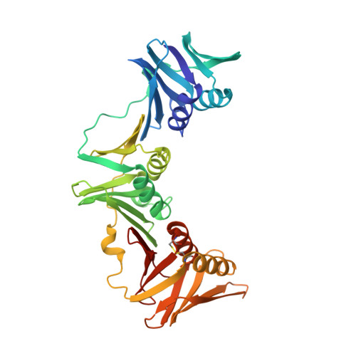
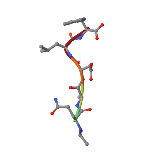
Structural and Thermodynamic Dissection of Linear Motif Recognition by the E. coli Sliding Clamp

Yin, Z.Kelso, M.J.Beck, J.L.Oakley, A.J.

(2013) J Med Chem 56: 8665-8673

  • DOI: https://doi.org/10.1021/jm401118f
  • Primary Citation of Related Structures:  
    4K3K, 4K3L, 4K3M, 4K3O, 4K3P, 4K3Q, 4K3R, 4K3S

  • PubMed Abstract: 

    Protein-protein interactions based on linear motif (LM) recognition play roles in many cell regulatory processes. The E. coli sliding clamp is a protein mediator of replisome formation, which uses a common surface pocket composed of two subsites (I and II) to interact with LMs in multiple binding partners. A structural and thermodynamic dissection of sliding clamp-LM recognition has been performed, providing support for a sequential binding model. According to the model, a hydrophobic C-terminal LM dipeptide submotif acts as an anchor to establish initial contacts within subsite I, and this is followed by formation of a stabilizing hydrogen-bonding network between the flanking LM residues and subsite II. Differential solvation/desolvation during positioning of the submotifs is proposed as a driver for the sequential binding. Our model provides general insights into linear motif recognition and should guide the design of small-molecule inhibitors of the E. coli sliding clamp, an emerging antibacterial target.


  • Organizational Affiliation

    School of Chemistry, University of Wollongong , Northfields Avenue, Wollongong 2522, NSW, Australia.


Macromolecules
Find similar proteins by:  (by identity cutoff)  |  3D Structure
Entity ID: 1
MoleculeChains Sequence LengthOrganismDetailsImage
DNA polymerase III subunit beta
A, B
366Escherichia coli K-12Mutation(s): 0 
Gene Names: DnaN
EC: 2.7.7.7
UniProt
Find proteins for P0A988 (Escherichia coli (strain K12))
Explore P0A988 
Go to UniProtKB:  P0A988
Entity Groups  
Sequence Clusters30% Identity50% Identity70% Identity90% Identity95% Identity100% Identity
UniProt GroupP0A988
Sequence Annotations
Expand
  • Reference Sequence

Find similar proteins by:  Sequence   |   3D Structure  

Entity ID: 2
MoleculeChains Sequence LengthOrganismDetailsImage
(ACE)QADLFC [auth E]6N/AMutation(s): 0 
Sequence Annotations
Expand
  • Reference Sequence
Small Molecules
Ligands 5 Unique
IDChains Name / Formula / InChI Key2D Diagram3D Interactions
PG4
Query on PG4

Download Ideal Coordinates CCD File 
Q [auth B]TETRAETHYLENE GLYCOL
C8 H18 O5
UWHCKJMYHZGTIT-UHFFFAOYSA-N
PGE
Query on PGE

Download Ideal Coordinates CCD File 
I [auth A],
R [auth B],
T [auth B]
TRIETHYLENE GLYCOL
C6 H14 O4
ZIBGPFATKBEMQZ-UHFFFAOYSA-N
PEG
Query on PEG

Download Ideal Coordinates CCD File 
D [auth A],
L [auth B],
M [auth B],
U [auth B]
DI(HYDROXYETHYL)ETHER
C4 H10 O3
MTHSVFCYNBDYFN-UHFFFAOYSA-N
EDO
Query on EDO

Download Ideal Coordinates CCD File 
K [auth A],
S [auth B]
1,2-ETHANEDIOL
C2 H6 O2
LYCAIKOWRPUZTN-UHFFFAOYSA-N
CA
Query on CA

Download Ideal Coordinates CCD File 
E [auth A]
F [auth A]
G [auth A]
H [auth A]
J [auth A]
E [auth A],
F [auth A],
G [auth A],
H [auth A],
J [auth A],
N [auth B],
O [auth B],
P [auth B]
CALCIUM ION
Ca
BHPQYMZQTOCNFJ-UHFFFAOYSA-N
Experimental Data & Validation

Experimental Data

  • Method: X-RAY DIFFRACTION
  • Resolution: 2.00 Å
  • R-Value Free: 0.248 
  • R-Value Work: 0.197 
  • R-Value Observed: 0.200 
  • Space Group: P 1 21 1
Unit Cell:
Length ( Å )Angle ( ˚ )
a = 79.985α = 90
b = 67.246β = 113.78
c = 81.432γ = 90
Software Package:
Software NamePurpose
SCALEPACKdata scaling
REFMACrefinement
PDB_EXTRACTdata extraction
MAR345dtbdata collection
HKL-2000data reduction
MOLREPphasing

Structure Validation

View Full Validation Report


Entry History 

Deposition Data

Revision History  (Full details and data files)

  • Version 1.0: 2013-05-01
    Type: Initial release
  • Version 1.1: 2014-01-15
    Changes: Database references
  • Version 1.2: 2023-11-08
    Changes: Data collection, Database references, Derived calculations, Refinement description