4K3H

Immunoglobulin lambda variable domain L5(L89S) fluorogen activationg protein in complex with malachite green


Experimental Data Snapshot

  • Method: X-RAY DIFFRACTION
  • Resolution: 2.45 Å
  • R-Value Free: 0.225 
  • R-Value Work: 0.194 
  • R-Value Observed: 0.196 

wwPDB Validation   3D Report Full Report


Ligand Structure Quality Assessment 


This is version 1.2 of the entry. See complete history


Literature

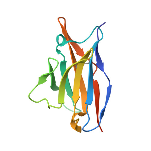
Malachite Green Mediates Homodimerization of Antibody VL Domains to Form a Fluorescent Ternary Complex with Singular Symmetric Interfaces.

Szent-Gyorgyi, C.Stanfield, R.L.Andreko, S.Dempsey, A.Ahmed, M.Capek, S.Waggoner, A.S.Wilson, I.A.Bruchez, M.P.

(2013) J Mol Biol 425: 4595-4613

  • DOI: https://doi.org/10.1016/j.jmb.2013.08.014
  • Primary Citation of Related Structures:  
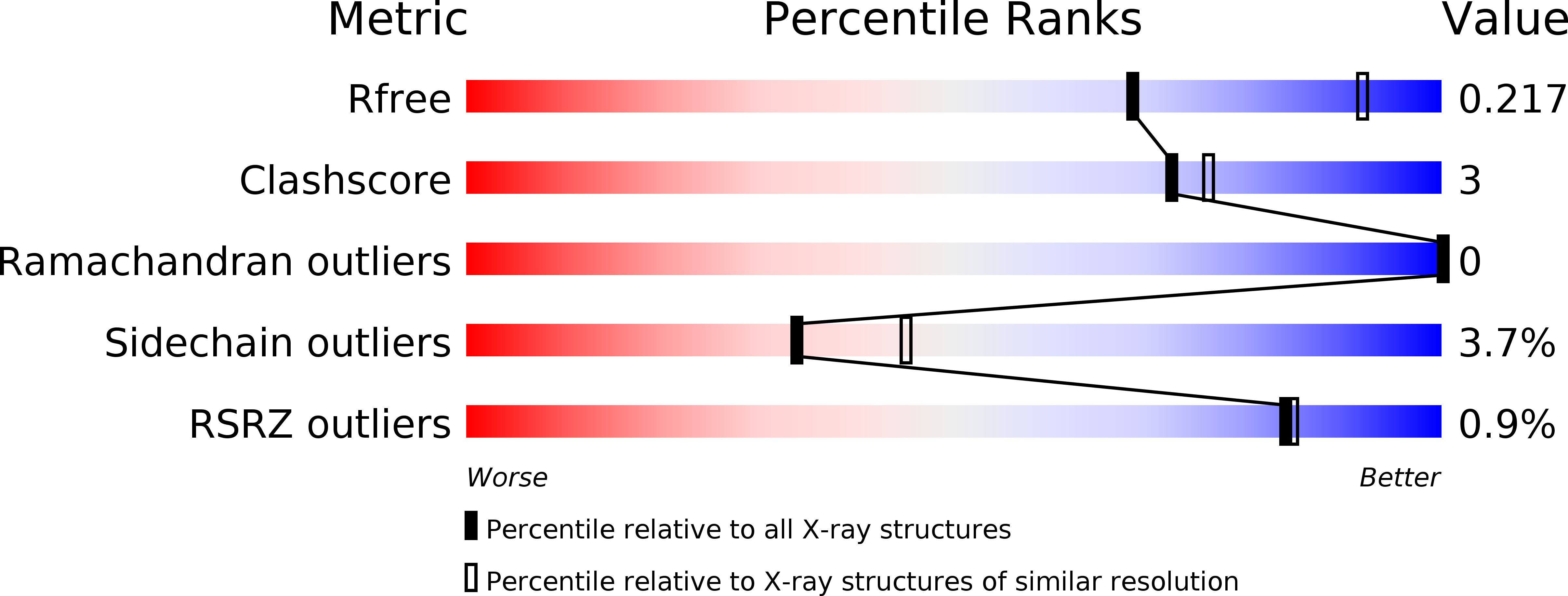
    4K3G, 4K3H

  • PubMed Abstract: 

    We report that a symmetric small-molecule ligand mediates the assembly of antibody light chain variable domains (VLs) into a correspondent symmetric ternary complex with novel interfaces. The L5* fluorogen activating protein is a VL domain that binds malachite green (MG) dye to activate intense fluorescence. Crystallography of liganded L5* reveals a 2:1 protein:ligand complex with inclusive C2 symmetry, where MG is almost entirely encapsulated between an antiparallel arrangement of the two VL domains. Unliganded L5* VL domains crystallize as a similar antiparallel VL/VL homodimer. The complementarity-determining regions are spatially oriented to form novel VL/VL and VL/ligand interfaces that tightly constrain a propeller conformer of MG. Binding equilibrium analysis suggests highly cooperative assembly to form a very stable VL/MG/VL complex, such that MG behaves as a strong chemical inducer of dimerization. Fusion of two VL domains into a single protein tightens MG binding over 1000-fold to low picomolar affinity without altering the large binding enthalpy, suggesting that bonding interactions with ligand and restriction of domain movements make independent contributions to binding. Fluorescence activation of a symmetrical fluorogen provides a selection mechanism for the isolation and directed evolution of ternary complexes where unnatural symmetric binding interfaces are favored over canonical antibody interfaces. As exemplified by L5*, these self-reporting complexes may be useful as modulators of protein association or as high-affinity protein tags and capture reagents.


  • Organizational Affiliation

    Molecular Biosensor and Imaging Center, Carnegie Mellon University, Pittsburgh, PA 15213, USA. Electronic address: css@andrew.cmu.edu.


Macromolecules
Find similar proteins by:  (by identity cutoff)  |  3D Structure
Entity ID: 1
MoleculeChains Sequence LengthOrganismDetailsImage
Immunoglobulin lambda variable domain L5(L89S)
A, B, C, D, E
A, B, C, D, E, F, G, H
118Homo sapiensMutation(s): 0 
Entity Groups  
Sequence Clusters30% Identity50% Identity70% Identity90% Identity95% Identity100% Identity
Sequence Annotations
Expand
  • Reference Sequence
Small Molecules
Ligands 3 Unique
IDChains Name / Formula / InChI Key2D Diagram3D Interactions
1OM
Query on 1OM

Download Ideal Coordinates CCD File 
I [auth A],
O [auth D],
R [auth E],
W [auth G]
4-{bis[4-(dimethylamino)phenyl]methyl}phenol
C23 H26 N2 O
ZTXGOUSDKAXJJB-UHFFFAOYSA-N
MAN
Query on MAN

Download Ideal Coordinates CCD File 
AA [auth H],
Q [auth D],
U [auth E]
alpha-D-mannopyranose
C6 H12 O6
WQZGKKKJIJFFOK-PQMKYFCFSA-N
GOL
Query on GOL

Download Ideal Coordinates CCD File 
J [auth B]
K [auth B]
L [auth B]
M [auth B]
N [auth C]
J [auth B],
K [auth B],
L [auth B],
M [auth B],
N [auth C],
P [auth D],
S [auth E],
T [auth E],
V [auth F],
X [auth G],
Y [auth H],
Z [auth H]
GLYCEROL
C3 H8 O3
PEDCQBHIVMGVHV-UHFFFAOYSA-N
Binding Affinity Annotations 
IDSourceBinding Affinity
1OM Binding MOAD:  4K3H Kd: 0.09 (nM) from 1 assay(s)
PDBBind:  4K3H Kd: 140 (nM) from 1 assay(s)
Experimental Data & Validation

Experimental Data

  • Method: X-RAY DIFFRACTION
  • Resolution: 2.45 Å
  • R-Value Free: 0.225 
  • R-Value Work: 0.194 
  • R-Value Observed: 0.196 
  • Space Group: P 43
Unit Cell:
Length ( Å )Angle ( ˚ )
a = 94.11α = 90
b = 94.11β = 90
c = 175.8γ = 90
Software Package:
Software NamePurpose
Blu-Icedata collection
PHASERphasing
PHENIXrefinement
HKL-2000data reduction
HKL-2000data scaling

Structure Validation

View Full Validation Report


Ligand Structure Quality Assessment 


Entry History 

Deposition Data

Revision History  (Full details and data files)

  • Version 1.0: 2013-10-09
    Type: Initial release
  • Version 1.1: 2013-11-13
    Changes: Database references
  • Version 1.2: 2020-07-29
    Type: Remediation
    Reason: Carbohydrate remediation
    Changes: Data collection, Derived calculations, Structure summary