4K2R

Structural basis for activation of ZAP-70 by phosphorylation of the SH2-kinase linker


Experimental Data Snapshot

  • Method: X-RAY DIFFRACTION
  • Resolution: 3.00 Å
  • R-Value Free: 0.287 
  • R-Value Work: 0.209 
  • R-Value Observed: 0.213 

wwPDB Validation   3D Report Full Report


Ligand Structure Quality Assessment 


This is version 1.2 of the entry. See complete history


Literature

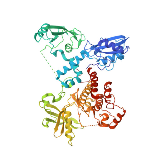
Structural Basis for Activation of ZAP-70 by Phosphorylation of the SH2-Kinase Linker.

Yan, Q.Barros, T.Visperas, P.R.Deindl, S.Kadlecek, T.A.Weiss, A.Kuriyan, J.

(2013) Mol Cell Biol 33: 2188-2201

  • DOI: https://doi.org/10.1128/MCB.01637-12
  • Primary Citation of Related Structures:  
    4K2R

  • PubMed Abstract: 

    Serial activation of the tyrosine kinases Lck and ZAP-70 initiates signaling downstream of the T cell receptor. We previously reported the structure of an autoinhibited ZAP-70 variant in which two regulatory tyrosine residues (315 and 319) in the SH2-kinase linker were replaced by phenylalanine. We now present a crystal structure of ZAP-70 in which Tyr 315 and Tyr 319 are not mutated, leading to the recognition of a five-residue sequence register error in the SH2-kinase linker of the original crystallographic model. The revised model identifies distinct roles for these two tyrosines. As seen in a recently reported structure of the related tyrosine kinase Syk, Tyr 315 of ZAP-70 is part of a hydrophobic interface between the regulatory apparatus and the kinase domain, and the integrity of this interface would be lost upon engagement of doubly phosphorylated peptides by the SH2 domains. Tyr 319 is not necessarily dislodged by SH2 engagement, which activates ZAP-70 only ∼5-fold in vitro. In contrast, phosphorylation by Lck activates ZAP-70 ∼100-fold. This difference is due to the ability of Tyr 319 to suppress ZAP-70 activity even when the SH2 domains are dislodged from the kinase domain, providing stringent control of ZAP-70 activity downstream of Lck.


  • Organizational Affiliation

    Department of Molecular and Cell Biology, University of California, Berkeley, CA, USA.


Macromolecules
Find similar proteins by:  (by identity cutoff)  |  3D Structure
Entity ID: 1
MoleculeChains Sequence LengthOrganismDetailsImage
Tyrosine-protein kinase ZAP-70614Homo sapiensMutation(s): 1 
Gene Names: ZAP70SRK
EC: 2.7.10.2
UniProt & NIH Common Fund Data Resources
Find proteins for P43403 (Homo sapiens)
Explore P43403 
Go to UniProtKB:  P43403
PHAROS:  P43403
GTEx:  ENSG00000115085 
Entity Groups  
Sequence Clusters30% Identity50% Identity70% Identity90% Identity95% Identity100% Identity
UniProt GroupP43403
Sequence Annotations
Expand
  • Reference Sequence
Experimental Data & Validation

Experimental Data

  • Method: X-RAY DIFFRACTION
  • Resolution: 3.00 Å
  • R-Value Free: 0.287 
  • R-Value Work: 0.209 
  • R-Value Observed: 0.213 
  • Space Group: P 1
Unit Cell:
Length ( Å )Angle ( ˚ )
a = 48.53α = 106.1
b = 53.15β = 93.26
c = 68.96γ = 104.4
Software Package:
Software NamePurpose
Blu-Icedata collection
MOLREPphasing
REFMACrefinement
MOSFLMdata reduction
SCALAdata scaling

Structure Validation

View Full Validation Report


Ligand Structure Quality Assessment 


Entry History 

Deposition Data

Revision History  (Full details and data files)

  • Version 1.0: 2013-05-08
    Type: Initial release
  • Version 1.1: 2013-05-22
    Changes: Database references
  • Version 1.2: 2024-02-28
    Changes: Data collection, Database references, Derived calculations